6Z3X | pdb_00006z3x

Crystal structure of the designed antibody DesAb-anti-HSA-P1


Experimental Data Snapshot

  • Method: X-RAY DIFFRACTION
  • Resolution: 1.74 Å
  • R-Value Free: 
    0.220 (Depositor), 0.228 (DCC) 
  • R-Value Work: 
    0.193 (Depositor), 0.197 (DCC) 
  • R-Value Observed: 
    0.194 (Depositor) 

Starting Model: experimental
View more details

wwPDB Validation 3D Report Full Report

Validation slider image for 6Z3X

This is version 1.3 of the entry. See complete history

Literature

Fragment-based computational design of antibodies targeting structured epitopes.

Aguilar Rangel, M.Bedwell, A.Costanzi, E.Taylor, R.J.Russo, R.Bernardes, G.J.L.Ricagno, S.Frydman, J.Vendruscolo, M.Sormanni, P.

(2022) Sci Adv 8: eabp9540-eabp9540

  • DOI: https://doi.org/10.1126/sciadv.abp9540
  • Primary Citation Related Structures: 
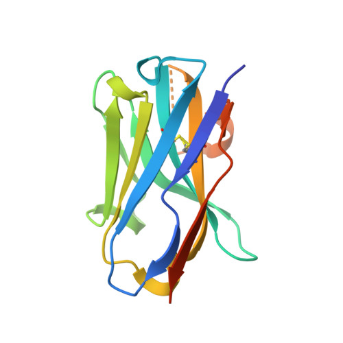
    6Z3X

  • PubMed Abstract: 

    De novo design methods hold the promise of reducing the time and cost of antibody discovery while enabling the facile and precise targeting of predetermined epitopes. Here, we describe a fragment-based method for the combinatorial design of antibody binding loops and their grafting onto antibody scaffolds. We designed and tested six single-domain antibodies targeting different epitopes on three antigens, including the receptor-binding domain of the SARS-CoV-2 spike protein. Biophysical characterization showed that all designs are stable and bind their intended targets with affinities in the nanomolar range without in vitro affinity maturation. We further discuss how a high-resolution input antigen structure is not required, as similar predictions are obtained when the input is a crystal structure or a computer-generated model. This computational procedure, which readily runs on a laptop, provides a starting point for the rapid generation of lead antibodies binding to preselected epitopes.


  • Organizational Affiliation
    • Centre for Misfolding Diseases, Yusuf Hamied Department of Chemistry, University of Cambridge, Cambridge CB2 1EW, UK.

Macromolecule Content 

  • Total Structure Weight: 30.98 kDa 
  • Atom Count: 2,037 
  • Modeled Residue Count: 240 
  • Deposited Residue Count: 274 
  • Unique protein chains: 1

Macromolecules

Find similar proteins by:|  3D Structure
Entity ID: 1
MoleculeChains  Sequence LengthOrganismDetailsImage
DesAb-anti-HSA-P1
A, B
137synthetic constructMutation(s): 0 

Small Molecules

Ligands 3 Unique
IDChains Name / Formula / InChI Key2D Diagram3D Interactions
CAC

Query on CAC


Download:Ideal Coordinates CCD File
C [auth A]
D [auth B]
E [auth B]
F [auth B]
H [auth B]
C [auth A],
D [auth B],
E [auth B],
F [auth B],
H [auth B],
I [auth B]
CACODYLATE ION
C2 H6 As O2
OGGXGZAMXPVRFZ-UHFFFAOYSA-M
IMD

Query on IMD


Download:Ideal Coordinates CCD File
G [auth B]IMIDAZOLE
C3 H5 N2
RAXXELZNTBOGNW-UHFFFAOYSA-O
NA

Query on NA


Download:Ideal Coordinates CCD File
J [auth B]SODIUM ION
Na
FKNQFGJONOIPTF-UHFFFAOYSA-N

Experimental Data & Validation

Experimental Data

  • Method: X-RAY DIFFRACTION
  • Resolution: 1.74 Å
  • R-Value Free:  0.220 (Depositor), 0.228 (DCC) 
  • R-Value Work:  0.193 (Depositor), 0.197 (DCC) 
  • R-Value Observed: 0.194 (Depositor) 
Space Group: P 21 21 21
Unit Cell:
Length ( Å )Angle ( ˚ )
a = 40.718α = 90
b = 52.411β = 90
c = 99.176γ = 90
Software Package:
Software NamePurpose
PHENIXrefinement
PHENIXrefinement
DIALSdata reduction
Aimlessdata scaling
PHASERphasing

Structure Validation

View Full Validation Report


Entry History 

Deposition Data

Revision History  (Full details and data files)

  • Version 1.0: 2021-03-31
    Type: Initial release
  • Version 1.1: 2022-11-23
    Changes: Database references
  • Version 1.2: 2024-01-31
    Changes: Data collection, Refinement description
  • Version 1.3: 2024-11-13
    Changes: Structure summary