Cryo-EM reveals structural breaks in a patient-derived amyloid fibril from systemic AL amyloidosis.
Radamaker, L., Baur, J., Huhn, S., Haupt, C., Hegenbart, U., Schonland, S., Bansal, A., Schmidt, M., Fandrich, M.(2021) Nat Commun 12: 875-875
- PubMed: 33558536 Search on PubMedSearch on PubMed Central
- DOI: https://doi.org/10.1038/s41467-021-21126-2
- Primary Citation Related Structures:
6Z1I, 6Z1O - PubMed Abstract:
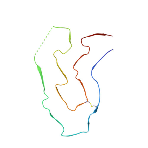
Systemic AL amyloidosis is a debilitating and potentially fatal disease that arises from the misfolding and fibrillation of immunoglobulin light chains (LCs). The disease is patient-specific with essentially each patient possessing a unique LC sequence. In this study, we present two ex vivo fibril structures of a λ3 LC. The fibrils were extracted from the explanted heart of a patient (FOR005) and consist of 115-residue fibril proteins, mainly from the LC variable domain. The fibril structures imply that a 180° rotation around the disulfide bond and a major unfolding step are necessary for fibrils to form. The two fibril structures show highly similar fibril protein folds, differing in only a 12-residue segment. Remarkably, the two structures do not represent separate fibril morphologies, as they can co-exist at different z-axial positions within the same fibril. Our data imply the presence of structural breaks at the interface of the two structural forms.
- Institute of Protein Biochemistry, Ulm University, Ulm, Germany.
Organizational Affiliation: