6YYP | pdb_00006yyp

Structure of Cathepsin S in complex with Compound 2


Experimental Data Snapshot

  • Method: X-RAY DIFFRACTION
  • Resolution: 2.05 Å
  • R-Value Free: 
    0.188 (Depositor), 0.194 (DCC) 
  • R-Value Work: 
    0.141 (Depositor), 0.152 (DCC) 

Starting Model: experimental
View more details

wwPDB Validation 3D Report Full Report

Validation slider image for 6YYP

Ligand Structure Quality Assessment 


This is version 1.3 of the entry. See complete history

Literature

Highly Selective Sub-Nanomolar Cathepsin S Inhibitors by Merging Fragment Binders with Nitrile Inhibitors.

Schade, M.Merla, B.Lesch, B.Wagener, M.Timmermanns, S.Pletinckx, K.Hertrampf, T.

(2020) J Med Chem 63: 11801-11808

  • DOI: https://doi.org/10.1021/acs.jmedchem.0c00949
  • Primary Citation Related Structures: 
    6YYN, 6YYO, 6YYP, 6YYQ, 6YYR

  • PubMed Abstract: 

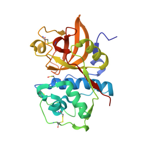
    Pharmacological inhibition of cathepsin S (CatS) allows for a specific modulation of the adaptive immune system and many major diseases. Here, we used NMR fragment screening and crystal structure-aided merging to synthesize novel, highly selective CatS inhibitors with picomolar enzymatic Ki values and nanomolar functional activity in human Raji cells. Noncovalent fragment hits revealed binding hotspots, while the covalent inhibitor structure-activity relationship enabled efficient potency optimization.


  • Organizational Affiliation
    • Grünenthal GmbH, Zieglerstr. 6, 52078 Aachen, Germany.

Macromolecule Content 

  • Total Structure Weight: 51.21 kDa 
  • Atom Count: 3,993 
  • Modeled Residue Count: 442 
  • Deposited Residue Count: 450 
  • Unique protein chains: 1

Macromolecules

Find similar proteins by:|  3D Structure
Entity ID: 1
MoleculeChains  Sequence LengthOrganismDetailsImage
Cathepsin SA [auth AAA],
B [auth BBB]
225Homo sapiensMutation(s): 0 
Gene Names: CTSS
EC: 3.4.22.27
UniProt & NIH Common Fund Data Resources
Find proteins for P25774 (Homo sapiens)
Explore P25774 
Go to UniProtKB:  P25774
PHAROS:  P25774
GTEx:  ENSG00000163131 
Entity Groups
Sequence Clusters30% Identity50% Identity70% Identity90% Identity95% Identity100% Identity
UniProt GroupP25774
Sequence Annotations
Expand
Reference Sequence

Small Molecules

Ligands 4 Unique
IDChains Name / Formula / InChI Key2D Diagram3D Interactions
Q1H
(Subject of Investigation/LOI)

Query on Q1H


Download:Ideal Coordinates CCD File
E [auth AAA]1-(furan-2-ylmethyl)-5-(trifluoromethyl)benzimidazol-2-amine
C13 H10 F3 N3 O
QYVPLFHAOLRMBY-UHFFFAOYSA-N
SIN

Query on SIN


Download:Ideal Coordinates CCD File
F [auth BBB]SUCCINIC ACID
C4 H6 O4
KDYFGRWQOYBRFD-UHFFFAOYSA-N
MLA

Query on MLA


Download:Ideal Coordinates CCD File
G [auth BBB],
H [auth BBB]
MALONIC ACID
C3 H4 O4
OFOBLEOULBTSOW-UHFFFAOYSA-N
ACT

Query on ACT


Download:Ideal Coordinates CCD File
C [auth AAA]
D [auth AAA]
I [auth BBB]
J [auth BBB]
K [auth BBB]
C [auth AAA],
D [auth AAA],
I [auth BBB],
J [auth BBB],
K [auth BBB],
L [auth BBB]
ACETATE ION
C2 H3 O2
QTBSBXVTEAMEQO-UHFFFAOYSA-M
Modified Residues  1 Unique
IDChains TypeFormula2D DiagramParent
CSO
Query on CSO
A [auth AAA],
B [auth BBB]
L-PEPTIDE LINKINGC3 H7 N O3 SCYS

Experimental Data & Validation

Experimental Data

  • Method: X-RAY DIFFRACTION
  • Resolution: 2.05 Å
  • R-Value Free:  0.188 (Depositor), 0.194 (DCC) 
  • R-Value Work:  0.141 (Depositor), 0.152 (DCC) 
Space Group: H 3 2
Unit Cell:
Length ( Å )Angle ( ˚ )
a = 118.689α = 90
b = 118.689β = 90
c = 205.586γ = 120
Software Package:
Software NamePurpose
REFMACrefinement
MOSFLMdata reduction
SCALAdata scaling
REFMACphasing

Structure Validation

View Full Validation Report


Ligand Structure Quality Assessment 


Entry History 

Deposition Data

Revision History  (Full details and data files)

  • Version 1.0: 2021-05-12
    Type: Initial release
  • Version 1.1: 2022-11-23
    Changes: Database references, Derived calculations
  • Version 1.2: 2024-05-01
    Changes: Data collection, Refinement description
  • Version 1.3: 2024-10-16
    Changes: Structure summary