6YRA | pdb_00006yra

Crystal structure of ATP-dependent caprolactamase from Pseudomonas jessenii


Experimental Data Snapshot

  • Method: X-RAY DIFFRACTION
  • Resolution: 4.00 Å
  • R-Value Free: 
    0.381 (Depositor), 0.375 (DCC) 
  • R-Value Work: 
    0.239 (Depositor) 
  • R-Value Observed: 
    0.246 (Depositor) 

Starting Models: experimental
View more details

wwPDB Validation 3D Report Full Report

Validation slider image for 6YRA

This is version 1.3 of the entry. See complete history

Literature

Catalytic and structural properties of ATP-dependent caprolactamase from Pseudomonas jessenii.

Marjanovic, A.Rozeboom, H.J.de Vries, M.S.Mayer, C.Otzen, M.Wijma, H.J.Janssen, D.B.

(2021) Proteins 89: 1079-1098

  • DOI: https://doi.org/10.1002/prot.26082
  • Primary Citation Related Structures: 
    6YRA

  • PubMed Abstract: 

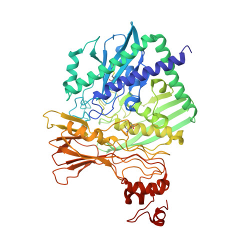
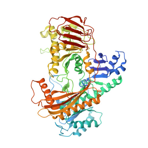
    Caprolactamase is the first enzyme in the caprolactam degradation pathway of Pseudomonas jessenii. It is composed of two subunits (CapA and CapB) and sequence-related to other ATP-dependent enzymes involved in lactam hydrolysis, like 5-oxoprolinases and hydantoinases. Low sequence similarity also exists with ATP-dependent acetone- and acetophenone carboxylases. The caprolactamase was produced in Escherichia coli, isolated by His-tag affinity chromatography, and subjected to functional and structural studies. Activity toward caprolactam required ATP and was dependent on the presence of bicarbonate in the assay buffer. The hydrolysis product was identified as 6-aminocaproic acid. Quantum mechanical modeling indicated that the hydrolysis of caprolactam was highly disfavored (ΔG 0 '= 23 kJ/mol), which explained the ATP dependence. A crystal structure showed that the enzyme exists as an (αβ) 2 tetramer and revealed an ATP-binding site in CapA and a Zn-coordinating site in CapB. Mutations in the ATP-binding site of CapA (D11A and D295A) significantly reduced product formation. Mutants with substitutions in the metal binding site of CapB (D41A, H99A, D101A, and H124A) were inactive and less thermostable than the wild-type enzyme. These residues proved to be essential for activity and on basis of the experimental findings we propose possible mechanisms for ATP-dependent lactam hydrolysis.


  • Organizational Affiliation
    • Biotransformation and Biocatalysis, Groningen Biomolecular Sciences and Biotechnology Institute (GBB), University of Groningen, Groningen, The Netherlands.

Macromolecule Content 

  • Total Structure Weight: 276.57 kDa 
  • Atom Count: 19,265 
  • Modeled Residue Count: 2,540 
  • Deposited Residue Count: 2,554 
  • Unique protein chains: 2

Macromolecules

Find similar proteins by:|  3D Structure
Entity ID: 1
MoleculeChains  Sequence LengthOrganismDetailsImage
HydantoinaseA [auth B],
C [auth D]
581Pseudomonas jesseniiMutation(s): 0 
Gene Names: CRX42_01180
EC: 3.5.2
UniProt
Find proteins for A0A2W0FH34 (Pseudomonas jessenii)
Explore A0A2W0FH34 
Go to UniProtKB:  A0A2W0FH34
Entity Groups
Sequence Clusters30% Identity50% Identity70% Identity90% Identity95% Identity100% Identity
UniProt GroupA0A2W0FH34
Sequence Annotations
Expand
Reference Sequence
Find similar proteins by:|  3D Structure
Entity ID: 2
MoleculeChains  Sequence LengthOrganismDetailsImage
5-oxoprolinaseB [auth A],
D [auth C]
696Pseudomonas jesseniiMutation(s): 0 
Gene Names: CRX42_01175
EC: 3.5.2
UniProt
Find proteins for A0A2W0EVE0 (Pseudomonas jessenii)
Explore A0A2W0EVE0 
Go to UniProtKB:  A0A2W0EVE0
Entity Groups
Sequence Clusters30% Identity50% Identity70% Identity90% Identity95% Identity100% Identity
UniProt GroupA0A2W0EVE0
Sequence Annotations
Expand
Reference Sequence

Experimental Data & Validation

Experimental Data

  • Method: X-RAY DIFFRACTION
  • Resolution: 4.00 Å
  • R-Value Free:  0.381 (Depositor), 0.375 (DCC) 
  • R-Value Work:  0.239 (Depositor) 
  • R-Value Observed: 0.246 (Depositor) 
Space Group: P 21 21 2
Unit Cell:
Length ( Å )Angle ( ˚ )
a = 195.377α = 90
b = 167.367β = 90
c = 87.965γ = 90
Software Package:
Software NamePurpose
PHENIXrefinement
XDSdata reduction
Aimlessdata scaling
PHASERphasing
PDB_EXTRACTdata extraction

Structure Validation

View Full Validation Report


Entry History 

Deposition Data

Revision History  (Full details and data files)

  • Version 1.0: 2021-04-21
    Type: Initial release
  • Version 1.1: 2021-08-18
    Changes: Database references
  • Version 1.2: 2024-01-24
    Changes: Data collection, Refinement description
  • Version 1.3: 2024-11-06
    Changes: Structure summary