6YP1 | pdb_00006yp1

HiCel7B unliganded


Experimental Data Snapshot

  • Method: X-RAY DIFFRACTION
  • Resolution: 1.20 Å
  • R-Value Free: 
    0.135 (Depositor), 0.135 (DCC) 
  • R-Value Work: 
    0.117 (Depositor), 0.117 (DCC) 

Starting Model: experimental
View more details

wwPDB Validation 3D Report Full Report

Validation slider image for 6YP1

Ligand Structure Quality Assessment 


This is version 2.2 of the entry. See complete history

Literature

Glycosylated cyclophellitol-derived activity-based probes and inhibitors for cellulases.

de Boer, C.McGregor, N.G.S.Peterse, E.Schroder, S.P.Florea, B.I.Jiang, J.Reijngoud, J.Ram, A.F.J.van Wezel, G.P.van der Marel, G.A.Codee, J.D.C.Overkleeft, H.S.Davies, G.J.

(2020) RSC Chem Biol 1: 148-155

  • DOI: https://doi.org/10.1039/d0cb00045k
  • Primary Citation Related Structures: 
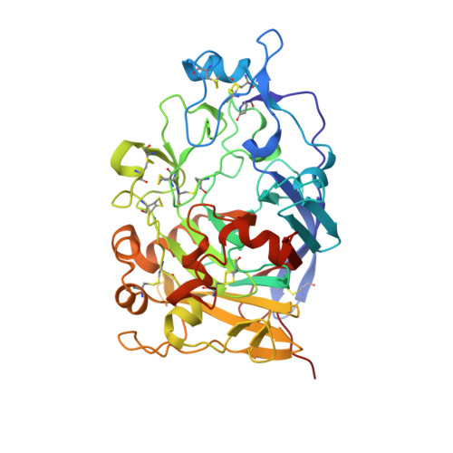
    6YOZ, 6YP1

  • PubMed Abstract: 

    Cellulases and related β-1,4-glucanases are essential components of lignocellulose-degrading enzyme mixtures. The detection of β-1,4-glucanase activity typically relies on monitoring the breakdown of purified lignocellulose-derived substrates or synthetic chromogenic substrates, limiting the activities which can be detected and complicating the tracing of activity back to specific components within complex enzyme mixtures. As a tool for the rapid detection and identification of β-1,4-glucanases, a series of glycosylated cyclophellitol inhibitors mimicking β-1,4-glucan oligosaccharides have been synthesised. These compounds are highly efficient inhibitors of HiCel7B, a well-known GH7 endo -β-1,4-glucanase. An elaborated activity-based probe facilitated the direct detection and identification of β-1,4-glucanases within a complex fungal secretome without any detectable cross-reactivity with β-d-glucosidases. These probes and inhibitors add valuable new capacity to the growing toolbox of cyclophellitol-derived probes for the activity-based profiling of biomass-degrading enzymes.


  • Organizational Affiliation
    • Leiden Institute of Chemistry, Leiden University Einsteinweg 55 2300 RA Leiden The Netherlands h.s.overkleeft@chem.leidenuniv.nl.

Macromolecule Content 

  • Total Structure Weight: 46.44 kDa 
  • Atom Count: 3,651 
  • Modeled Residue Count: 402 
  • Deposited Residue Count: 402 
  • Unique protein chains: 1

Macromolecules

Find similar proteins by:|  3D Structure
Entity ID: 1
MoleculeChains  Sequence LengthOrganismDetailsImage
Endoglucanase 1A [auth AAA]402Mycothermus thermophilusMutation(s): 0 
Gene Names: CEL7B
EC: 3.2.1.4
UniProt
Find proteins for P56680 (Humicola insolens)
Explore P56680 
Go to UniProtKB:  P56680
Entity Groups
Sequence Clusters30% Identity50% Identity70% Identity90% Identity95% Identity100% Identity
UniProt GroupP56680
Glycosylation
Glycosylation Sites: 2
Sequence Annotations
Expand
Reference Sequence

Small Molecules

Ligands 4 Unique
IDChains Name / Formula / InChI Key2D Diagram3D Interactions
NAG

Query on NAG


Download:Ideal Coordinates CCD File
H [auth AAA],
R [auth AAA]
2-acetamido-2-deoxy-beta-D-glucopyranose
C8 H15 N O6
OVRNDRQMDRJTHS-FMDGEEDCSA-N
SO4

Query on SO4


Download:Ideal Coordinates CCD File
C [auth AAA]
D [auth AAA]
E [auth AAA]
I [auth AAA]
J [auth AAA]
C [auth AAA],
D [auth AAA],
E [auth AAA],
I [auth AAA],
J [auth AAA],
K [auth AAA],
N [auth AAA],
P [auth AAA],
Q [auth AAA]
SULFATE ION
O4 S
QAOWNCQODCNURD-UHFFFAOYSA-L
GOL

Query on GOL


Download:Ideal Coordinates CCD File
B [auth AAA],
F [auth AAA],
L [auth AAA],
M [auth AAA],
O [auth AAA]
GLYCEROL
C3 H8 O3
PEDCQBHIVMGVHV-UHFFFAOYSA-N
ACM

Query on ACM


Download:Ideal Coordinates CCD File
G [auth AAA]ACETAMIDE
C2 H5 N O
DLFVBJFMPXGRIB-UHFFFAOYSA-N
Modified Residues  1 Unique
IDChains TypeFormula2D DiagramParent
PCA
Query on PCA
A [auth AAA]L-PEPTIDE LINKINGC5 H7 N O3GLN

Experimental Data & Validation

Experimental Data

  • Method: X-RAY DIFFRACTION
  • Resolution: 1.20 Å
  • R-Value Free:  0.135 (Depositor), 0.135 (DCC) 
  • R-Value Work:  0.117 (Depositor), 0.117 (DCC) 
Space Group: P 42 21 2
Unit Cell:
Length ( Å )Angle ( ˚ )
a = 92.31α = 90
b = 92.31β = 90
c = 100.81γ = 90
Software Package:
Software NamePurpose
REFMACrefinement
xia2data reduction
xia2data scaling
PHASERphasing

Structure Validation

View Full Validation Report


Ligand Structure Quality Assessment 


Entry History 

& Funding Information

Deposition Data


Funding OrganizationLocationGrant Number
Biotechnology and Biological Sciences Research Council (BBSRC)United KingdomBB/R001162/1

Revision History  (Full details and data files)

  • Version 1.0: 2020-09-16
    Type: Initial release
  • Version 2.0: 2021-10-13
    Changes: Advisory, Atomic model, Data collection, Database references, Derived calculations, Structure summary
  • Version 2.1: 2024-01-24
    Changes: Data collection, Derived calculations, Refinement description
  • Version 2.2: 2024-11-06
    Changes: Structure summary