


Substrate Engagement and Catalytic Mechanisms of N-Acetylglucosaminyltransferase V
Darby, J.F., Gilio, A.K., Piniello, B., Roth, C., Blagova, E., Rovira, C., Hubbard, R.E., Davies, G.J., Wu, L.(2020) ACS Catal
Experimental Data Snapshot
Starting Model: experimental
View more details
(2020) ACS Catal
Entity ID: 1 | |||||
|---|---|---|---|---|---|
| Molecule | Chains | Sequence Length | Organism | Details | Image |
| Alpha-1,6-mannosylglycoprotein 6-beta-N-acetylglucosaminyltransferase A | A [auth AAA], B [auth BBB] | 515 | Homo sapiens | Mutation(s): 0 Gene Names: MGAT5, GGNT5 EC: 2.4.1.155 | 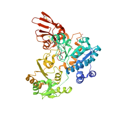 |
UniProt & NIH Common Fund Data Resources | |||||
PHAROS: Q09328 GTEx: ENSG00000152127 | |||||
Entity Groups | |||||
| Sequence Clusters | 30% Identity50% Identity70% Identity90% Identity95% Identity100% Identity | ||||
| UniProt Group | Q09328 | ||||
Glycosylation | |||||
| Glycosylation Sites: 1 | Go to GlyGen: Q09328-1 | ||||
Sequence AnnotationsExpand | |||||
Reference Sequence | |||||
Entity ID: 2 | |||||
|---|---|---|---|---|---|
| Molecule | Chains | Length | 2D Diagram | Glycosylation | D Interactions |
| 2-acetamido-2-deoxy-beta-D-glucopyranose-(1-2)-alpha-D-mannopyranose-(1-3)-[2-acetamido-2-deoxy-beta-D-glucopyranose-(1-2)-alpha-D-mannopyranose-(1-6)]alpha-D-mannopyranose | C [auth A], D [auth B] | 5 |  | N/A | |
Glycosylation Resources | |||||
GlyTouCan: G19266OP GlyCosmos: G19266OP GlyGen: G19266OP | |||||
| Ligands 3 Unique | |||||
|---|---|---|---|---|---|
| ID | Chains | Name / Formula / InChI Key | 2D Diagram | 3D Interactions | |
| NAG (Subject of Investigation/LOI) Download:Ideal Coordinates CCD File | H [auth AAA], R [auth BBB] | 2-acetamido-2-deoxy-beta-D-glucopyranose C8 H15 N O6 OVRNDRQMDRJTHS-FMDGEEDCSA-N |  | ||
| SO4 Download:Ideal Coordinates CCD File | AA [auth BBB] I [auth AAA] J [auth AAA] K [auth AAA] L [auth AAA] | SULFATE ION O4 S QAOWNCQODCNURD-UHFFFAOYSA-L |  | ||
| EDO Download:Ideal Coordinates CCD File | E [auth AAA] F [auth AAA] G [auth AAA] N [auth BBB] O [auth BBB] | 1,2-ETHANEDIOL C2 H6 O2 LYCAIKOWRPUZTN-UHFFFAOYSA-N |  | ||
| Length ( Å ) | Angle ( ˚ ) |
|---|---|
| a = 46.28 | α = 108.81 |
| b = 67.37 | β = 92.2 |
| c = 91.05 | γ = 106.72 |
| Software Name | Purpose |
|---|---|
| REFMAC | refinement |
| xia2 | data reduction |
| xia2 | data scaling |
| REFMAC | phasing |
| Funding Organization | Location | Grant Number |
|---|---|---|
| European Research Council (ERC) | United Kingdom | ErC-2012-AdG-322942 |


