Structural Characterization of TssL from Acinetobacter baumannii: a Key Component of the Type VI Secretion System.
Ruiz, F.M., Lopez, J., Ferrara, C.G., Santillana, E., Espinosa, Y.R., Feldman, M.F., Romero, A.(2020) J Bacteriol 202
- PubMed: 32571965 Search on PubMedSearch on PubMed Central
- DOI: https://doi.org/10.1128/JB.00210-20
- Primary Citation Related Structures:
6Y4R - PubMed Abstract:
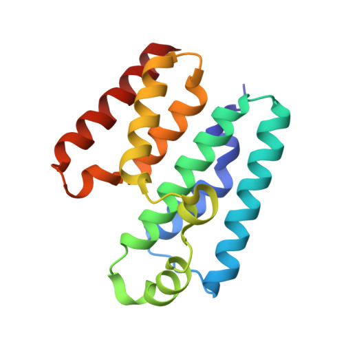
The type VI secretion system (T6SS) is a complex molecular nanomachine used by Gram-negative bacteria to deliver diverse effectors into adjacent cells. A membrane complex (MC) anchors this transport system to the bacterial cell wall. One of the proteins forming the MC is TssL, a cytoplasmic protein bound to the inner membrane through a single transmembrane helix. Here, we report the structure of the cytoplasmic N-terminal region of TssL from Acinetobacter baumannii , a bacterium encoding in a single locus a secretion system that is a special case among other T6SSs. The protein structure, consisting of two antiparallel alpha-helical bundles connected by a short loop, reveals several interesting particularities compared with homologous proteins from other organisms. In addition, we demonstrate the structural significance of residues Asp98 and Glu99, which are strongly conserved among T6SS-encoding Gram-negative bacteria. Mutations in these two residues strongly impact protein dynamics, expression, and functionality. Our results improve our understanding of the T6SS of A. baumannii , which remains largely understudied compared with that of other pathogens. IMPORTANCE Several Acinetobacter species carry one functional type VI secretion system (T6SS). The T6SS is encoded in a single locus containing 16 conserved genes, most of which code for proteins essential to T6SS activity. One of these key components is TssL, a cytoplasmic protein bound to the inner membrane. Despite its importance and its particular characteristics, the structure of T6SS in A. baumannii remains understudied. Here, we present structural, in silico , and in vivo studies of TssL, highlighting the importance of two well-conserved residues and improving our understanding of this secretion system in this bacterium.
- Chemical and Physical Biology, Centro de Investigaciones Biológicas Margarita Salas, Madrid, Spain fruiz@cib.csic.es romero@cib.csic.es.
Organizational Affiliation: