6Y1X | pdb_00006y1x

X-ray structure of the radical SAM protein NifB, a key nitrogenase maturating enzyme


Experimental Data Snapshot

  • Method: X-RAY DIFFRACTION
  • Resolution: 1.95 Å
  • R-Value Free: 
    0.212 (Depositor), 0.210 (DCC) 
  • R-Value Work: 
    0.177 (Depositor), 0.178 (DCC) 
  • R-Value Observed: 
    0.179 (Depositor) 

wwPDB Validation 3D Report Full Report

Validation slider image for 6Y1X

This is version 1.2 of the entry. See complete history

Literature

Structural Insights into the Mechanism of the Radical SAM Carbide Synthase NifB, a Key Nitrogenase Cofactor Maturating Enzyme.

Fajardo, A.S.Legrand, P.Paya-Tormo, L.A.Martin, L.Pellicer Marti Nez, M.T.Echavarri-Erasun, C.Vernede, X.Rubio, L.M.Nicolet, Y.

(2020) J Am Chem Soc 142: 11006-11012

  • DOI: https://doi.org/10.1021/jacs.0c02243
  • Primary Citation Related Structures: 
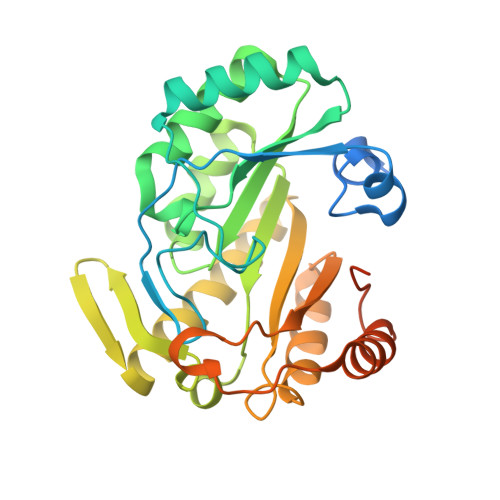
    6Y1X

  • PubMed Abstract: 

    Nitrogenase is a key player in the global nitrogen cycle, as it catalyzes the reduction of dinitrogen into ammonia. The active site of the nitrogenase MoFe protein corresponds to a [MoFe 7 S 9 C-( R )-homocitrate] species designated FeMo-cofactor, whose biosynthesis and insertion requires the action of over a dozen maturation proteins provided by the NIF (for NI trogen F ixation) assembly machinery. Among them, the radical SAM protein NifB plays an essential role, concomitantly inserting a carbide ion and coupling two [Fe 4 S 4 ] clusters to form a [Fe 8 S 9 C] precursor called NifB-co. Here we report on the X-ray structure of NifB from Methanotrix thermoacetophila at 1.95 Å resolution in a state pending the binding of one [Fe 4 S 4 ] cluster substrate. The overall NifB architecture indicates that this enzyme has a single SAM binding site, which at this stage is occupied by cysteine residue 62. The structure reveals a unique ligand binding mode for the K1-cluster involving cysteine residues 29 and 128 in addition to histidine 42 and glutamate 65. The latter, together with cysteine 62, belongs to a loop inserted in the active site, likely protecting the already present [Fe 4 S 4 ] clusters. These two residues regulate the sequence of events, controlling SAM dual reactivity and preventing unwanted radical-based chemistry before the K2 [Fe 4 S 4 ] cluster substrate is loaded into the protein. The location of the K1-cluster, too far away from the SAM binding site, supports a mechanism in which the K2-cluster is the site of methylation.


  • Organizational Affiliation
    • Centro de Biotecnologı́a y Genómica de Plantas, Universidad Politécnica de Madrid, Instituto Nacional de Investigación y Tecnologı́a Agraria y Alimentaria, Pozuelo de Alarcón, 28223 Madrid, Spain.

Macromolecule Content 

  • Total Structure Weight: 72.55 kDa 
  • Atom Count: 4,519 
  • Modeled Residue Count: 504 
  • Deposited Residue Count: 618 
  • Unique protein chains: 1

Macromolecules

Find similar proteins by:|  3D Structure
Entity ID: 1
MoleculeChains  Sequence LengthOrganismDetailsImage
Radical SAM domain protein
A, B
309Methanothrix thermoacetophila PTMutation(s): 0 
Gene Names: Mthe_0422
UniProt
Find proteins for A0B690 (Methanothrix thermoacetophila (strain DSM 6194 / JCM 14653 / NBRC 101360 / PT))
Explore A0B690 
Go to UniProtKB:  A0B690
Entity Groups
Sequence Clusters30% Identity50% Identity70% Identity90% Identity95% Identity100% Identity
UniProt GroupA0B690
Sequence Annotations
Expand
Reference Sequence

Small Molecules

Ligands 3 Unique
IDChains Name / Formula / InChI Key2D Diagram3D Interactions
SF4
(Subject of Investigation/LOI)

Query on SF4


Download:Ideal Coordinates CCD File
C [auth A],
D [auth A],
K [auth B],
L [auth B]
IRON/SULFUR CLUSTER
Fe4 S4
LJBDFODJNLIPKO-UHFFFAOYSA-N
SO4

Query on SO4


Download:Ideal Coordinates CCD File
E [auth A],
F [auth A],
H [auth B],
I [auth B],
J [auth B]
SULFATE ION
O4 S
QAOWNCQODCNURD-UHFFFAOYSA-L
CL

Query on CL


Download:Ideal Coordinates CCD File
G [auth A],
M [auth B]
CHLORIDE ION
Cl
VEXZGXHMUGYJMC-UHFFFAOYSA-M

Experimental Data & Validation

Experimental Data

  • Method: X-RAY DIFFRACTION
  • Resolution: 1.95 Å
  • R-Value Free:  0.212 (Depositor), 0.210 (DCC) 
  • R-Value Work:  0.177 (Depositor), 0.178 (DCC) 
  • R-Value Observed: 0.179 (Depositor) 
Space Group: P 1
Unit Cell:
Length ( Å )Angle ( ˚ )
a = 40.18α = 78.81
b = 48.68β = 87.73
c = 80.38γ = 89.94
Software Package:
Software NamePurpose
XDSdata reduction
Aimlessdata scaling
PHASERphasing
SHELXDEphasing
PHENIXrefinement
PDB_EXTRACTdata extraction

Structure Validation

View Full Validation Report


Entry History 

& Funding Information

Deposition Data


Funding OrganizationLocationGrant Number
French National Research AgencyFranceANR-17-EURE-0003
French National Research AgencyFranceANR-10-INBS-05-02
French National Research AgencyFranceANR-15-IDEX-02
Spanish Ministry of Science, Innovation, and UniversitiesSpain2017-88475-R

Revision History  (Full details and data files)

  • Version 1.0: 2020-06-17
    Type: Initial release
  • Version 1.1: 2020-07-01
    Changes: Database references
  • Version 1.2: 2024-05-15
    Changes: Data collection, Database references