6Y1U | pdb_00006y1u

Mycobacterium tuberculosis FtsZ-GDP in complex with 4-hydroxycoumarin


Experimental Data Snapshot

  • Method: X-RAY DIFFRACTION
  • Resolution: 1.68 Å
  • R-Value Free: 
    0.197 (Depositor), 0.197 (DCC) 
  • R-Value Work: 
    0.168 (Depositor), 0.170 (DCC) 
  • R-Value Observed: 
    0.170 (Depositor) 

Starting Model: experimental
View more details

wwPDB Validation 3D Report Full Report

Validation slider image for 6Y1U

Ligand Structure Quality Assessment 


This is version 1.2 of the entry. See complete history

Literature

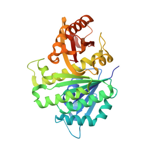
Conformational Flexibility of A Highly Conserved Helix Controls Cryptic Pocket Formation in FtsZ.

Alnami, A.Norton, R.S.Pena, H.P.Haider, S.Kozielski, F.

(2021) J Mol Biology 433: 167061-167061

  • DOI: https://doi.org/10.1016/j.jmb.2021.167061
  • Primary Citation Related Structures: 
    6Y1U, 6Y1V, 6YM1, 6YM9

  • PubMed Abstract: 

    Mycobacterium tuberculosis is responsible for more than 1.6 million deaths each year. One potential antibacterial target in M. tuberculosis is filamentous temperature sensitive protein Z (FtsZ), which is the bacterial homologue of mammalian tubulin, a validated cancer target. M. tuberculosis FtsZ function is essential, with its inhibition leading to arrest of cell division, elongation of the bacterial cell and eventual cell death. However, the development of potent inhibitors against FtsZ has been a challenge owing to the lack of structural information. Here we report multiple crystal structures of M. tuberculosis FtsZ in complex with a coumarin analogue. The 4-hydroxycoumarin binds exclusively to two novel cryptic pockets in nucleotide-free FtsZ, but not to the binary FtsZ-GTP or GDP complexes. Our findings provide a detailed understanding of the molecular basis for cryptic pocket formation, controlled by the conformational flexibility of the H7 helix, and thus reveal an important structural and mechanistic rationale for coumarin antibacterial activity.


  • Organizational Affiliation
    • Department of Pharmaceutical and Biological Chemistry, School of Pharmacy, University College London, 29-39 Brunswick Square, London WC1N 1AX, UK.

Macromolecule Content 

  • Total Structure Weight: 66.57 kDa 
  • Atom Count: 4,888 
  • Modeled Residue Count: 598 
  • Deposited Residue Count: 628 
  • Unique protein chains: 1

Macromolecules

Find similar proteins by:|  3D Structure
Entity ID: 1
MoleculeChains  Sequence LengthOrganismDetailsImage
Cell division protein FtsZ
A, B
314Mycobacterium tuberculosis CDC1551Mutation(s): 0 
Gene Names: ftsZMT2209
UniProt
Find proteins for P9WN95 (Mycobacterium tuberculosis (strain ATCC 25618 / H37Rv))
Explore P9WN95 
Go to UniProtKB:  P9WN95
Entity Groups
Sequence Clusters30% Identity50% Identity70% Identity90% Identity95% Identity100% Identity
UniProt GroupP9WN95
Sequence Annotations
Expand
Reference Sequence

Small Molecules

Ligands 3 Unique
IDChains Name / Formula / InChI Key2D Diagram3D Interactions
GDP

Query on GDP


Download:Ideal Coordinates CCD File
K [auth A]GUANOSINE-5'-DIPHOSPHATE
C10 H15 N5 O11 P2
QGWNDRXFNXRZMB-UUOKFMHZSA-N
4HC

Query on 4HC


Download:Ideal Coordinates CCD File
Y [auth B],
Z [auth B]
4-HYDROXY-2H-CHROMEN-2-ONE
C9 H6 O3
VXIXUWQIVKSKSA-UHFFFAOYSA-N
DMS

Query on DMS


Download:Ideal Coordinates CCD File
C [auth A]
D [auth A]
E [auth A]
F [auth A]
G [auth A]
C [auth A],
D [auth A],
E [auth A],
F [auth A],
G [auth A],
H [auth A],
I [auth A],
J [auth A],
L [auth B],
M [auth B],
N [auth B],
O [auth B],
P [auth B],
Q [auth B],
R [auth B],
S [auth B],
T [auth B],
U [auth B],
V [auth B],
W [auth B],
X [auth B]
DIMETHYL SULFOXIDE
C2 H6 O S
IAZDPXIOMUYVGZ-UHFFFAOYSA-N

Experimental Data & Validation

Experimental Data

  • Method: X-RAY DIFFRACTION
  • Resolution: 1.68 Å
  • R-Value Free:  0.197 (Depositor), 0.197 (DCC) 
  • R-Value Work:  0.168 (Depositor), 0.170 (DCC) 
  • R-Value Observed: 0.170 (Depositor) 
Space Group: P 65
Unit Cell:
Length ( Å )Angle ( ˚ )
a = 88.13α = 90
b = 88.13β = 90
c = 176.783γ = 120
Software Package:
Software NamePurpose
PHENIXrefinement
iMOSFLMdata reduction
SCALAdata scaling
PHASERphasing

Structure Validation

View Full Validation Report


Ligand Structure Quality Assessment 


Entry History 

Deposition Data

Revision History  (Full details and data files)

  • Version 1.0: 2021-06-09
    Type: Initial release
  • Version 1.1: 2021-06-16
    Changes: Database references, Structure summary
  • Version 1.2: 2024-01-24
    Changes: Data collection, Database references, Refinement description