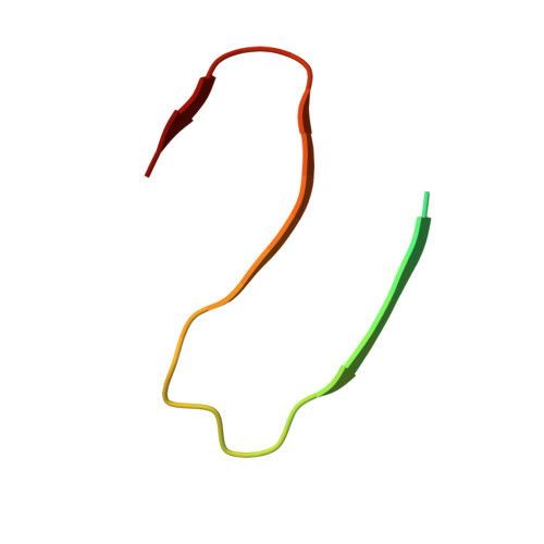
6Y1A

Amyloid fibril structure of islet amyloid polypeptide


Experimental Data Snapshot

  • Method: ELECTRON MICROSCOPY
  • Resolution: 4.20 Å
  • Aggregation State: FILAMENT 
  • Reconstruction Method: HELICAL 

wwPDB Validation   3D Report Full Report


This is version 1.1 of the entry. See complete history


Literature

Cryo-EM structure of islet amyloid polypeptide fibrils reveals similarities with amyloid-beta fibrils.

Roder, C.Kupreichyk, T.Gremer, L.Schafer, L.U.Pothula, K.R.Ravelli, R.B.G.Willbold, D.Hoyer, W.Schroder, G.F.

(2020) Nat Struct Mol Biol 27: 660-667

  • DOI: https://doi.org/10.1038/s41594-020-0442-4
  • Primary Citation of Related Structures:  
    6Y1A

  • PubMed Abstract: 

    Amyloid deposits consisting of fibrillar islet amyloid polypeptide (IAPP) in pancreatic islets are associated with beta-cell loss and have been implicated in type 2 diabetes (T2D). Here, we applied cryo-EM to reconstruct densities of three dominant IAPP fibril polymorphs, formed in vitro from synthetic human IAPP. An atomic model of the main polymorph, built from a density map of 4.2-Å resolution, reveals two S-shaped, intertwined protofilaments. The segment 21-NNFGAIL-27, essential for IAPP amyloidogenicity, forms the protofilament interface together with Tyr37 and the amidated C terminus. The S-fold resembles polymorphs of Alzheimer's disease (AD)-associated amyloid-β (Aβ) fibrils, which might account for the epidemiological link between T2D and AD and reports on IAPP-Aβ cross-seeding in vivo. The results structurally link the early-onset T2D IAPP genetic polymorphism (encoding Ser20Gly) with the AD Arctic mutation (Glu22Gly) of Aβ and support the design of inhibitors and imaging probes for IAPP fibrils.


  • Organizational Affiliation

    Institute of Biological Information Processing (IBI-7: Structural Biochemistry), Forschungszentrum Jülich, Jülich, Germany.


Macromolecules
Find similar proteins by:  (by identity cutoff)  |  3D Structure
Entity ID: 1
MoleculeChains Sequence LengthOrganismDetailsImage
Islet amyloid polypeptide
A, B, C, D, E
A, B, C, D, E, F, G, H, I, J, K, L, M, N, O, P
37Homo sapiensMutation(s): 0 
UniProt & NIH Common Fund Data Resources
Find proteins for P10997 (Homo sapiens)
Explore P10997 
Go to UniProtKB:  P10997
PHAROS:  P10997
GTEx:  ENSG00000121351 
Entity Groups  
Sequence Clusters30% Identity50% Identity70% Identity90% Identity95% Identity100% Identity
UniProt GroupP10997
Sequence Annotations
Expand
  • Reference Sequence
Small Molecules
Ligands 1 Unique
IDChains Name / Formula / InChI Key2D Diagram3D Interactions
NH2
Query on NH2

Download Ideal Coordinates CCD File 
AA [auth K]
BA [auth L]
CA [auth M]
DA [auth N]
EA [auth O]
AA [auth K],
BA [auth L],
CA [auth M],
DA [auth N],
EA [auth O],
FA [auth P],
Q [auth A],
R [auth B],
S [auth C],
T [auth D],
U [auth E],
V [auth F],
W [auth G],
X [auth H],
Y [auth I],
Z [auth J]
AMINO GROUP
H2 N
QGZKDVFQNNGYKY-UHFFFAOYSA-N
Experimental Data & Validation

Experimental Data

  • Method: ELECTRON MICROSCOPY
  • Resolution: 4.20 Å
  • Aggregation State: FILAMENT 
  • Reconstruction Method: HELICAL 
EM Software:
TaskSoftware PackageVersion
RECONSTRUCTIONRELION3.0.5
MODEL REFINEMENTPHENIX1.11

Structure Validation

View Full Validation Report


Entry History & Funding Information

Deposition Data


Funding OrganizationLocationGrant Number
European Research Council (ERC)European Union726368

Revision History  (Full details and data files)

  • Version 1.0: 2020-03-04
    Type: Initial release
  • Version 1.1: 2020-07-22
    Changes: Database references