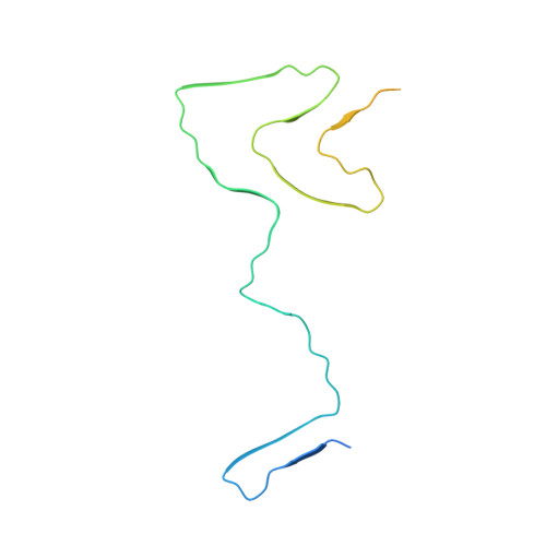
Structures of alpha-synuclein filaments from multiple system atrophy.
Schweighauser, M., Shi, Y., Tarutani, A., Kametani, F., Murzin, A.G., Ghetti, B., Matsubara, T., Tomita, T., Ando, T., Hasegawa, K., Murayama, S., Yoshida, M., Hasegawa, M., Scheres, S.H.W., Goedert, M.(2020) Nature 585: 464-469
- PubMed: 32461689
- DOI: https://doi.org/10.1038/s41586-020-2317-6
- Primary Citation of Related Structures:
6XYO, 6XYP, 6XYQ - PubMed Abstract:
Synucleinopathies, which include multiple system atrophy (MSA), Parkinson's disease, Parkinson's disease with dementia and dementia with Lewy bodies (DLB), are human neurodegenerative diseases 1 . Existing treatments are at best symptomatic. These diseases are characterized by the presence of, and believed to be caused by the formation of, filamentous inclusions of α-synuclein in brain cells 2,3 . However, the structures of α-synuclein filaments from the human brain are unknown. Here, using cryo-electron microscopy, we show that α-synuclein inclusions from the brains of individuals with MSA are made of two types of filament, each of which consists of two different protofilaments. In each type of filament, non-proteinaceous molecules are present at the interface of the two protofilaments. Using two-dimensional class averaging, we show that α-synuclein filaments from the brains of individuals with MSA differ from those of individuals with DLB, which suggests that distinct conformers or strains characterize specific synucleinopathies. As is the case with tau assemblies 4-9 , the structures of α-synuclein filaments extracted from the brains of individuals with MSA differ from those formed in vitro using recombinant proteins, which has implications for understanding the mechanisms of aggregate propagation and neurodegeneration in the human brain. These findings have diagnostic and potential therapeutic relevance, especially because of the unmet clinical need to be able to image filamentous α-synuclein inclusions in the human brain.
- MRC Laboratory of Molecular Biology, Cambridge, UK.
Organizational Affiliation:


