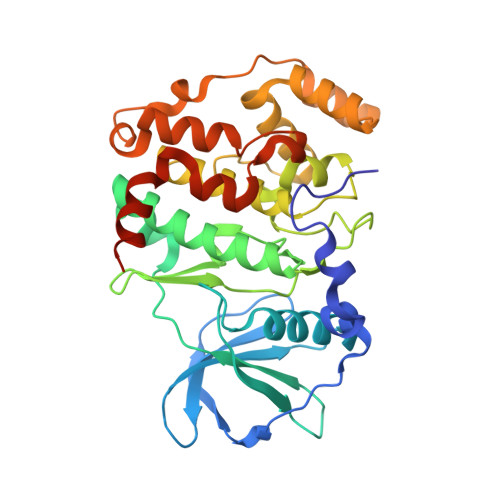
6XX7

Arabidopsis thaliana Casein Kinase 2 (CK2) alpha-1 crystal in complex with ANP


Experimental Data Snapshot

  • Method: X-RAY DIFFRACTION
  • Resolution: 2.40 Å
  • R-Value Free: 0.246 
  • R-Value Work: 0.195 
  • R-Value Observed: 0.198 

Starting Model: experimental
View more details

wwPDB Validation   3D Report Full Report


Ligand Structure Quality Assessment 


This is version 1.1 of the entry. See complete history


Literature

Crystal structure of Arabidopsis thaliana casein kinase 2 alpha 1.

Demulder, M.De Veylder, L.Loris, R.

(2020) Acta Crystallogr F Struct Biol Commun 76: 182-191

  • DOI: https://doi.org/10.1107/S2053230X20004537
  • Primary Citation of Related Structures:  
    6XX6, 6XX7, 6XX8, 6XX9

  • PubMed Abstract: 

    Casein kinase 2 (CK2) is a ubiquitous pleiotropic enzyme that is highly conserved across eukaryotic kingdoms. CK2 is singular amongst kinases as it is highly rigid and constitutively active. Arabidopsis thaliana is widely used as a model system in molecular plant research; the biological functions of A. thaliana CK2 are well studied in vivo and many of its substrates have been identified. Here, crystal structures of the α subunit of A. thaliana CK2 in three crystal forms and of its complex with the nonhydrolyzable ATP analog AMppNHp are presented. While the C-lobe of the enzyme is highly rigid, structural plasticity is observed for the N-lobe. Small but significant displacements within the active cleft are necessary in order to avoid steric clashes with the AMppNHp molecule. Binding of AMppNHp is influenced by a rigid-body motion of the N-lobe that was not previously recognized in maize CK2.


  • Organizational Affiliation

    Structural Biology Brussels, Vrije Universiteit Brussel, Pleinlaan 2, B-1050 Brussels, Belgium.


Macromolecules
Find similar proteins by:  (by identity cutoff)  |  3D Structure
Entity ID: 1
MoleculeChains Sequence LengthOrganismDetailsImage
Casein kinase II subunit alpha-1342Arabidopsis thalianaMutation(s): 0 
Gene Names: CKA1At5g67380K8K14.10
EC: 2.7.11.1
UniProt
Find proteins for Q08467 (Arabidopsis thaliana)
Explore Q08467 
Go to UniProtKB:  Q08467
Entity Groups  
Sequence Clusters30% Identity50% Identity70% Identity90% Identity95% Identity100% Identity
UniProt GroupQ08467
Sequence Annotations
Expand
  • Reference Sequence
Small Molecules
Ligands 5 Unique
IDChains Name / Formula / InChI Key2D Diagram3D Interactions
ANP (Subject of Investigation/LOI)
Query on ANP

Download Ideal Coordinates CCD File 
B [auth A]PHOSPHOAMINOPHOSPHONIC ACID-ADENYLATE ESTER
C10 H17 N6 O12 P3
PVKSNHVPLWYQGJ-KQYNXXCUSA-N
PGE
Query on PGE

Download Ideal Coordinates CCD File 
H [auth A]TRIETHYLENE GLYCOL
C6 H14 O4
ZIBGPFATKBEMQZ-UHFFFAOYSA-N
SO4
Query on SO4

Download Ideal Coordinates CCD File 
D [auth A]SULFATE ION
O4 S
QAOWNCQODCNURD-UHFFFAOYSA-L
CL
Query on CL

Download Ideal Coordinates CCD File 
E [auth A],
F [auth A],
G [auth A]
CHLORIDE ION
Cl
VEXZGXHMUGYJMC-UHFFFAOYSA-M
MG
Query on MG

Download Ideal Coordinates CCD File 
C [auth A]MAGNESIUM ION
Mg
JLVVSXFLKOJNIY-UHFFFAOYSA-N
Experimental Data & Validation

Experimental Data

  • Method: X-RAY DIFFRACTION
  • Resolution: 2.40 Å
  • R-Value Free: 0.246 
  • R-Value Work: 0.195 
  • R-Value Observed: 0.198 
  • Space Group: P 41 21 2
Unit Cell:
Length ( Å )Angle ( ˚ )
a = 60.56α = 90
b = 60.56β = 90
c = 228.6γ = 90
Software Package:
Software NamePurpose
PHASERphasing
PHENIXrefinement
XDSdata reduction
XDSdata scaling

Structure Validation

View Full Validation Report


Ligand Structure Quality Assessment 


Entry History & Funding Information

Deposition Data


Funding OrganizationLocationGrant Number
Research Foundation - Flanders (FWO)BelgiumG023616N
Research Foundation - Flanders (FWO)BelgiumG011420N

Revision History  (Full details and data files)

  • Version 1.0: 2020-04-15
    Type: Initial release
  • Version 1.1: 2024-01-24
    Changes: Data collection, Database references, Refinement description