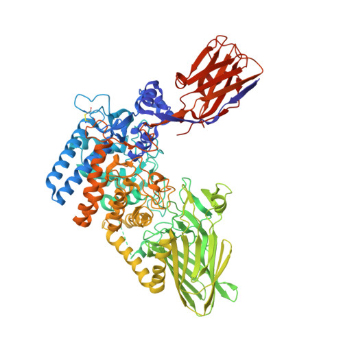
6XUX

Crystal structure of Megabody Mb-Nb207-cYgjK_NO


Experimental Data Snapshot

  • Method: X-RAY DIFFRACTION
  • Resolution: 1.90 Å
  • R-Value Free: 0.230 
  • R-Value Work: 0.189 
  • R-Value Observed: 0.191 

Starting Model: experimental
View more details

wwPDB Validation   3D Report Full Report


This is version 1.3 of the entry. See complete history


Literature

Megabodies expand the nanobody toolkit for protein structure determination by single-particle cryo-EM.

Uchanski, T.Masiulis, S.Fischer, B.Kalichuk, V.Lopez-Sanchez, U.Zarkadas, E.Weckener, M.Sente, A.Ward, P.Wohlkonig, A.Zogg, T.Remaut, H.Naismith, J.H.Nury, H.Vranken, W.Aricescu, A.R.Pardon, E.Steyaert, J.

(2021) Nat Methods 18: 60-68

  • DOI: https://doi.org/10.1038/s41592-020-01001-6
  • Primary Citation of Related Structures:  
    6QFA, 6XUX, 6XV8, 6XVI

  • PubMed Abstract: 

    Nanobodies are popular and versatile tools for structural biology. They have a compact single immunoglobulin domain organization, bind target proteins with high affinities while reducing their conformational heterogeneity and stabilize multi-protein complexes. Here we demonstrate that engineered nanobodies can also help overcome two major obstacles that limit the resolution of single-particle cryo-electron microscopy reconstructions: particle size and preferential orientation at the water-air interfaces. We have developed and characterized constructs, termed megabodies, by grafting nanobodies onto selected protein scaffolds to increase their molecular weight while retaining the full antigen-binding specificity and affinity. We show that the megabody design principles are applicable to different scaffold proteins and recognition domains of compatible geometries and are amenable for efficient selection from yeast display libraries. Moreover, we demonstrate that megabodies can be used to obtain three-dimensional reconstructions for membrane proteins that suffer from severe preferential orientation or are otherwise too small to allow accurate particle alignment.


  • Organizational Affiliation

    Structural Biology Brussels, Vrije Universiteit Brussel, VUB, Brussels, Belgium.


Macromolecules
Find similar proteins by:  (by identity cutoff)  |  3D Structure
Entity ID: 1
MoleculeChains Sequence LengthOrganismDetailsImage
Nanobody,Glucosidase YgjK,Glucosidase YgjK,Nanobody907Escherichia coli K-12Mutation(s): 0 
Gene Names: ygjKb3080JW3051
EC: 3.2.1
Membrane Entity: Yes 
UniProt
Find proteins for P42592 (Escherichia coli (strain K12))
Explore P42592 
Go to UniProtKB:  P42592
Entity Groups  
Sequence Clusters30% Identity50% Identity70% Identity90% Identity95% Identity100% Identity
UniProt GroupP42592
Sequence Annotations
Expand
  • Reference Sequence
Small Molecules
Ligands 1 Unique
IDChains Name / Formula / InChI Key2D Diagram3D Interactions
CA
Query on CA

Download Ideal Coordinates CCD File 
B [auth A]CALCIUM ION
Ca
BHPQYMZQTOCNFJ-UHFFFAOYSA-N
Experimental Data & Validation

Experimental Data

  • Method: X-RAY DIFFRACTION
  • Resolution: 1.90 Å
  • R-Value Free: 0.230 
  • R-Value Work: 0.189 
  • R-Value Observed: 0.191 
  • Space Group: P 21 21 21
Unit Cell:
Length ( Å )Angle ( ˚ )
a = 67.45α = 90
b = 84.46β = 90
c = 140.32γ = 90
Software Package:
Software NamePurpose
PHENIXrefinement
XDSdata reduction
XSCALEdata scaling
PHASERphasing

Structure Validation

View Full Validation Report


Entry History & Funding Information

Deposition Data


Funding OrganizationLocationGrant Number
Research Foundation - Flanders (FWO)Belgium--

Revision History  (Full details and data files)

  • Version 1.0: 2021-01-13
    Type: Initial release
  • Version 1.1: 2021-01-20
    Changes: Database references
  • Version 1.2: 2024-01-24
    Changes: Data collection, Database references, Refinement description
  • Version 1.3: 2024-10-23
    Changes: Structure summary