6XNY

Structure of RAG1 (R848M/E649V)-RAG2-DNA Strand Transfer Complex (Paired-Form)


Experimental Data Snapshot

  • Method: ELECTRON MICROSCOPY
  • Resolution: 2.90 Å
  • Aggregation State: PARTICLE 
  • Reconstruction Method: SINGLE PARTICLE 

Starting Model: experimental
View more details

wwPDB Validation   3D Report Full Report


This is version 1.3 of the entry. See complete history


Literature

Structural basis for the activation and suppression of transposition during evolution of the RAG recombinase.

Zhang, Y.Corbett, E.Wu, S.Schatz, D.G.

(2020) EMBO J 39: e105857-e105857

  • DOI: https://doi.org/10.15252/embj.2020105857
  • Primary Citation of Related Structures:  
    6XNX, 6XNY, 6XNZ

  • PubMed Abstract: 

    Jawed vertebrate adaptive immunity relies on the RAG1/RAG2 (RAG) recombinase, a domesticated transposase, for assembly of antigen receptor genes. Using an integration-activated form of RAG1 with methionine at residue 848 and cryo-electron microscopy, we determined structures that capture RAG engaged with transposon ends and U-shaped target DNA prior to integration (the target capture complex) and two forms of the RAG strand transfer complex that differ based on whether target site DNA is annealed or dynamic. Target site DNA base unstacking, flipping, and melting by RAG1 methionine 848 explain how this residue activates transposition, how RAG can stabilize sharp bends in target DNA, and why replacement of residue 848 by arginine during RAG domestication led to suppression of transposition activity. RAG2 extends a jawed vertebrate-specific loop to interact with target site DNA, and functional assays demonstrate that this loop represents another evolutionary adaptation acquired during RAG domestication to inhibit transposition. Our findings identify mechanistic principles of the final step in cut-and-paste transposition and the molecular and structural logic underlying the transformation of RAG from transposase to recombinase.


  • Organizational Affiliation

    Department of Immunobiology, Yale School of Medicine, New Haven, CT, USA.


Macromolecules

Find similar proteins by:  (by identity cutoff)  |  3D Structure
Entity ID: 1
MoleculeChains Sequence LengthOrganismDetailsImage
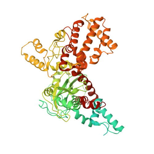
V(D)J recombination-activating protein 1
A, C
750Mus musculusMutation(s): 2 
Gene Names: Rag1
EC: 3.1 (PDB Primary Data), 2.3.2.27 (PDB Primary Data)
UniProt
Find proteins for P15919 (Mus musculus)
Explore P15919 
Go to UniProtKB:  P15919
Entity Groups  
Sequence Clusters30% Identity50% Identity70% Identity90% Identity95% Identity100% Identity
UniProt GroupP15919
Sequence Annotations
Expand
  • Reference Sequence
Find similar proteins by:  (by identity cutoff)  |  3D Structure
Entity ID: 2
MoleculeChains Sequence LengthOrganismDetailsImage
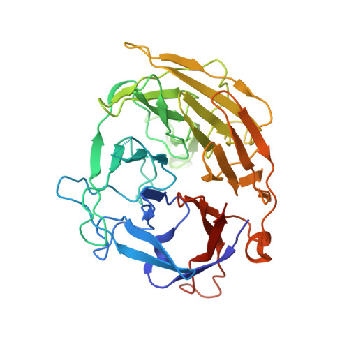
V(D)J recombination-activating protein 2
B, D
363Mus musculusMutation(s): 0 
Gene Names: Rag2Rag-2
UniProt
Find proteins for P21784 (Mus musculus)
Explore P21784 
Go to UniProtKB:  P21784
Entity Groups  
Sequence Clusters30% Identity50% Identity70% Identity90% Identity95% Identity100% Identity
UniProt GroupP21784
Sequence Annotations
Expand
  • Reference Sequence
Find similar nucleic acids by:  (by identity cutoff)  |  3D Structure
Entity ID: 3
MoleculeChains LengthOrganismImage
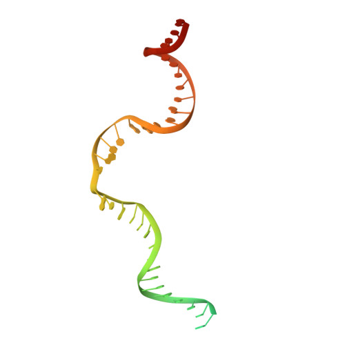
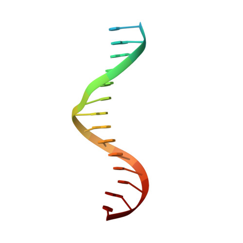
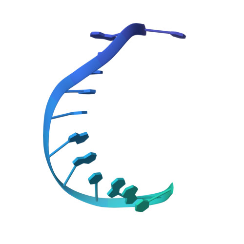
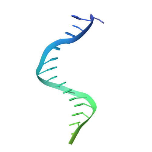
12RSS integration strand (55-mer)E [auth x]55Mus musculus
Sequence Annotations
Expand
  • Reference Sequence
Find similar nucleic acids by:  (by identity cutoff)  |  3D Structure
Entity ID: 4
MoleculeChains LengthOrganismImage
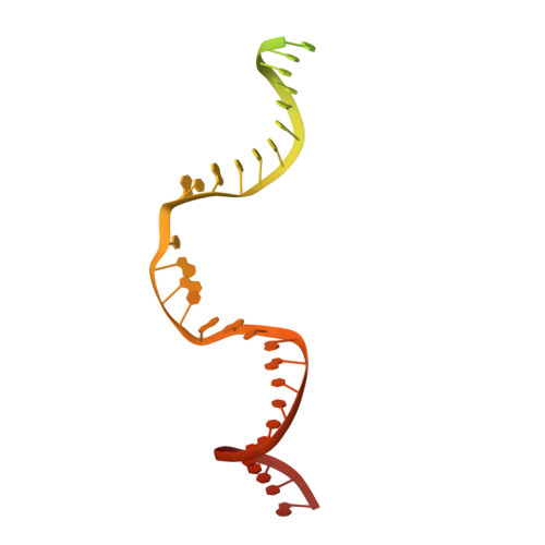
23RSS integration strand (66-mer)F [auth y]66Mus musculus
Sequence Annotations
Expand
  • Reference Sequence

Find similar nucleic acids by:  Sequence   |   3D Structure  

Entity ID: 5
MoleculeChains LengthOrganismImage
Flanking DNA top strand (16-mer)G [auth I],
H [auth J]
16Mus musculus
Sequence Annotations
Expand
  • Reference Sequence
Find similar nucleic acids by:  (by identity cutoff)  |  3D Structure
Entity ID: 6
MoleculeChains LengthOrganismImage
23RSS signal DNA top strand (45-mer)I [auth L]45Mus musculus
Sequence Annotations
Expand
  • Reference Sequence
Find similar nucleic acids by:  (by identity cutoff)  |  3D Structure
Entity ID: 7
MoleculeChains LengthOrganismImage
12RSS signal DNA top strand (34-mer)J [auth M]34Mus musculus
Sequence Annotations
Expand
  • Reference Sequence
Experimental Data & Validation

Experimental Data

  • Method: ELECTRON MICROSCOPY
  • Resolution: 2.90 Å
  • Aggregation State: PARTICLE 
  • Reconstruction Method: SINGLE PARTICLE 
EM Software:
TaskSoftware PackageVersion
MODEL REFINEMENTPHENIX1.17
RECONSTRUCTIONRELION3.0

Structure Validation

View Full Validation Report


Entry History & Funding Information

Deposition Data


Funding OrganizationLocationGrant Number
National Institutes of Health/National Institute of Diabetes and Digestive and Kidney Disease (NIH/NIDDK)United StatesR01 AI137079

Revision History  (Full details and data files)

  • Version 1.0: 2020-08-26
    Type: Initial release
  • Version 1.1: 2020-10-07
    Changes: Database references
  • Version 1.2: 2020-11-18
    Changes: Database references
  • Version 1.3: 2024-03-06
    Changes: Data collection, Database references, Refinement description