6XNJ | pdb_00006xnj

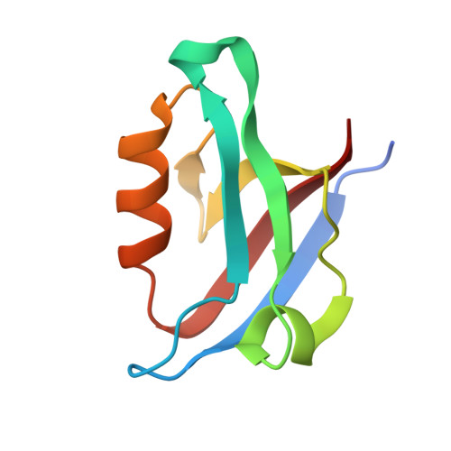
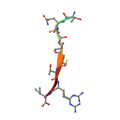
Crystal structure of the PDZ domain of human GOPC in complex with a peptide of E. coli O157:H7 str. Sakai effector NleG8


Experimental Data Snapshot

  • Method: X-RAY DIFFRACTION
  • Resolution: 1.85 Å
  • R-Value Free: 
    0.195 (Depositor), 0.193 (DCC) 
  • R-Value Work: 
    0.161 (Depositor), 0.162 (DCC) 
  • R-Value Observed: 
    0.163 (Depositor) 

Starting Model: experimental
View more details

wwPDB Validation 3D Report Full Report

Validation slider image for 6XNJ

This is version 1.3 of the entry. See complete history

Literature

Distinct Molecular Features of NleG Type 3 Secreted Effectors Allow for Different Roles during Citrobacter rodentium Infection in Mice.

Popov, G.Fiebig-Comyn, A.Syriste, L.Little, D.J.Skarina, T.Stogios, P.J.Birstonas, S.Coombes, B.K.Savchenko, A.

(2023) Infect Immun 91: e0050522-e0050522

  • DOI: https://doi.org/10.1128/iai.00505-22
  • Primary Citation Related Structures: 
    6XNJ

  • PubMed Abstract: 

    The NleGs are the largest family of type 3 secreted effectors in attaching and effacing (A/E) pathogens, such as enterohemorrhagic Escherichia coli (EHEC), enteropathogenic E. coli, and Citrobacter rodentium. NleG effectors contain a conserved C-terminal U-box domain acting as a ubiquitin protein ligase and target host proteins via a variable N-terminal portion. The specific roles of these effectors during infection remain uncertain. Here, we demonstrate that the three NleG effectors-NleG1 Cr , NleG7 Cr , and NleG8 Cr -encoded by C. rodentium DBS100 play distinct roles during infection in mice. Using individual nleG Cr knockout strains, we show that NleG7 Cr contributes to bacterial survival during enteric infection while NleG1 Cr promotes the expression of diarrheal symptoms and NleG8 Cr contributes to accelerated lethality in susceptible mice. Furthermore, the NleG8 Cr effector contains a C-terminal PDZ domain binding motif that enables interaction with the host protein GOPC. Both the PDZ domain binding motif and the ability to engage with host ubiquitination machinery via the intact U-box domain proved to be necessary for NleG8 Cr function, contributing to the observed phenotype during infection. We also establish that the PTZ binding motif in the EHEC NleG8 (NleG8 Ec ) effector, which shares 60% identity with NleG8 Cr , is engaged in interactions with human GOPC. The crystal structure of the NleG8 Ec C-terminal peptide in complex with the GOPC PDZ domain, determined to 1.85 Å, revealed a conserved interaction mode similar to that observed between GOPC and eukaryotic PDZ domain binding motifs. Despite these common features, nleG8 Ec does not complement the Δ nleG8 Cr phenotype during infection, revealing functional diversification between these NleG effectors.


  • Organizational Affiliation
    • Department of Microbiology, Immunology and Infectious Diseases, University of Calgary, Calgary, Alberta, Canada.

Macromolecule Content 

  • Total Structure Weight: 11.05 kDa 
  • Atom Count: 920 
  • Modeled Residue Count: 95 
  • Deposited Residue Count: 101 
  • Unique protein chains: 2

Macromolecules

Find similar proteins by:|  3D Structure
Entity ID: 1
MoleculeChains  Sequence LengthOrganismDetailsImage
Golgi-associated PDZ and coiled-coil motif-containing protein91Homo sapiensMutation(s): 0 
Gene Names: GOPCCALFIG
UniProt & NIH Common Fund Data Resources
Find proteins for Q9HD26 (Homo sapiens)
Explore Q9HD26 
Go to UniProtKB:  Q9HD26
GTEx:  ENSG00000047932 
Entity Groups
Sequence Clusters30% Identity50% Identity70% Identity90% Identity95% Identity100% Identity
UniProt GroupQ9HD26
Sequence Annotations
Expand
Reference Sequence
Find similar proteins by:  Sequence   |   3D Structure  
Entity ID: 2
MoleculeChains  Sequence LengthOrganismDetailsImage
NleG8 peptide10Escherichia coli O157:H7 str. SakaiMutation(s): 0 
Gene Names: NleG8
EC: 2.3.2.27
UniProt
Find proteins for Q8XAN6 (Escherichia coli O157:H7)
Explore Q8XAN6 
Go to UniProtKB:  Q8XAN6
Entity Groups
Sequence Clusters30% Identity50% Identity70% Identity90% Identity95% Identity100% Identity
UniProt GroupQ8XAN6
Sequence Annotations
Expand
Reference Sequence

Experimental Data & Validation

Experimental Data

  • Method: X-RAY DIFFRACTION
  • Resolution: 1.85 Å
  • R-Value Free:  0.195 (Depositor), 0.193 (DCC) 
  • R-Value Work:  0.161 (Depositor), 0.162 (DCC) 
  • R-Value Observed: 0.163 (Depositor) 
Space Group: P 32 2 1
Unit Cell:
Length ( Å )Angle ( ˚ )
a = 68.759α = 90
b = 68.759β = 90
c = 59.941γ = 120
Software Package:
Software NamePurpose
PHENIXrefinement
HKL-3000data reduction
HKL-3000data scaling
PHENIXphasing
PHENIXmodel building
Cootmodel building

Structure Validation

View Full Validation Report


Entry History 

& Funding Information

Deposition Data


Funding OrganizationLocationGrant Number
Canadian Institutes of Health Research (CIHR)Canada--
National Institutes of Health/National Institute Of Allergy and Infectious Diseases (NIH/NIAID)United States--

Revision History  (Full details and data files)

  • Version 1.0: 2020-08-12
    Type: Initial release
  • Version 1.1: 2023-10-18
    Changes: Data collection, Database references, Refinement description
  • Version 1.2: 2024-11-06
    Changes: Structure summary
  • Version 1.3: 2026-08-19
    Changes: Database references