


Crystal Structure of 2-hydroxyacyl CoA lyase (HACL) from Rhodospirillales bacterium URHD0017
Chou, A., Miller, M.D., Olmos Jr., J.L., Xu, W., Clomburg, J.M., Gonzalez, R., Philips Jr., G.N.To be published.
Experimental Data Snapshot
Starting Model: experimental
View more details
Entity ID: 1 | |||||
|---|---|---|---|---|---|
| Molecule | Chains | Sequence Length | Organism | Details | Image |
| 2-hydroxyacyl-CoA lyase 1 | 552 | Rhodospirillales bacterium URHD0017 | Mutation(s): 0 Gene Names: SAMN02990966_07839 | 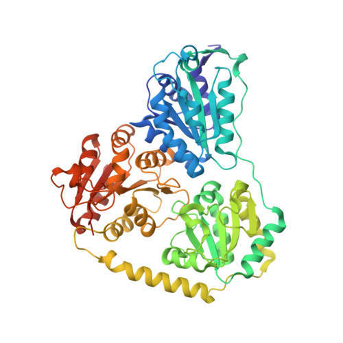 | |
| Ligands 4 Unique | |||||
|---|---|---|---|---|---|
| ID | Chains | Name / Formula / InChI Key | 2D Diagram | 3D Interactions | |
| TZD (Subject of Investigation/LOI) Download:Ideal Coordinates CCD File | D [auth A], H [auth B] | 2-{3-[(4-AMINO-2-METHYLPYRIMIDIN-5-YL)METHYL]-4-METHYL-2-OXO-2,3-DIHYDRO-1,3-THIAZOL-5-YL}ETHYL TRIHYDROGEN
DIPHOSPHATE C12 H18 N4 O8 P2 S ZGJUYGIRPQSCFA-UHFFFAOYSA-N |  | ||
| ADP (Subject of Investigation/LOI) Download:Ideal Coordinates CCD File | E [auth A], I [auth B] | ADENOSINE-5'-DIPHOSPHATE C10 H15 N5 O10 P2 XTWYTFMLZFPYCI-KQYNXXCUSA-N |  | ||
| MG (Subject of Investigation/LOI) Download:Ideal Coordinates CCD File | C [auth A], G [auth B] | MAGNESIUM ION Mg JLVVSXFLKOJNIY-UHFFFAOYSA-N |  | ||
| UNL Download:Ideal Coordinates CCD File | F [auth A], J [auth B] | Unknown ligand JLVVSXFLKOJNIY-UHFFFAOYSA-N | |||
| Length ( Å ) | Angle ( ˚ ) |
|---|---|
| a = 103.94 | α = 90 |
| b = 64.431 | β = 90.88 |
| c = 182.693 | γ = 90 |
| Software Name | Purpose |
|---|---|
| PDB_EXTRACT | data extraction |
| PHENIX | refinement |
| ARP/wARP | model building |
| PHASER | phasing |
| Aimless | data scaling |
| DIALS | data reduction |
| xia2 | data reduction |
| JBluIce-EPICS | data collection |
| Funding Organization | Location | Grant Number |
|---|---|---|
| National Science Foundation (NSF, United States) | United States | STC 1231306 |
| National Science Foundation (NSF, United States) | United States | CBET-1605999 |
| National Institutes of Health/National Institute of General Medical Sciences (NIH/NIGMS) | United States | R01 GM115261 |
| National Institutes of Health/National Cancer Institute (NIH/NCI) | United States | R01 CA217255 |
| National Science Foundation (NSF, United States) | United States | GRFP 1450681 |


