Phylogenetics-based identification and characterization of a superior 2,3-butanediol dehydrogenase for Zymomonas mobilis expression.
Subramanian, V., Lunin, V.V., Farmer, S.J., Alahuhta, M., Moore, K.T., Ho, A., Chaudhari, Y.B., Zhang, M., Himmel, M.E., Decker, S.R.(2020) Biotechnol Biofuels 13: 186-186
- PubMed: 33292448 Search on PubMedSearch on PubMed Central
- DOI: https://doi.org/10.1186/s13068-020-01820-x
- Primary Citation Related Structures:
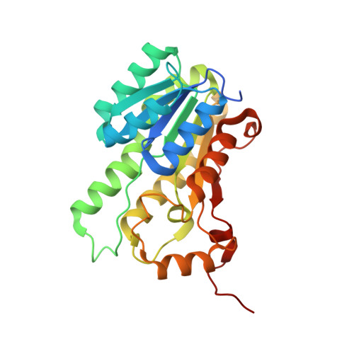
6VSP, 6XEW, 6XEX - PubMed Abstract:
Zymomonas mobilis has recently been shown to be capable of producing the valuable platform biochemical, 2,3-butanediol (2,3-BDO). Despite this capability, the production of high titers of 2,3-BDO is restricted by several physiological parameters. One such bottleneck involves the conversion of acetoin to 2,3-BDO, a step catalyzed by 2,3-butanediol dehydrogenase (Bdh). Several Bdh enzymes have been successfully expressed in Z. mobilis, although a highly active enzyme is yet to be identified for expression in this host. Here, we report the application of a phylogenetic approach to identify and characterize a superior Bdh, followed by validation of its structural attributes using a mutagenesis approach. Of the 11 distinct bdh genes that were expressed in Z. mobilis, crude extracts expressing Serratia marcescens Bdh (SmBdh) were found to have the highest activity (8.89 µmol/min/mg), when compared to other Bdh enzymes (0.34-2.87 µmol/min/mg). The SmBdh crystal structure was determined through crystallization with cofactor (NAD + ) and substrate (acetoin) molecules bound in the active site. Active SmBdh was shown to be a tetramer with the active site populated by a Gln247 residue contributed by the diagonally opposite subunit. SmBdh showed a more extensive supporting hydrogen-bond network in comparison to the other well-studied Bdh enzymes, which enables improved substrate positioning and substrate specificity. This protein also contains a short α6 helix, which provides more efficient entry and exit of molecules from the active site, thereby contributing to enhanced substrate turnover. Extending the α6 helix to mimic the lower activity Enterobacter cloacae (EcBdh) enzyme resulted in reduction of SmBdh function to nearly 3% of the total activity. In great contrast, reduction of the corresponding α6 helix of the EcBdh to mimic the SmBdh structure resulted in ~ 70% increase in its activity. This study has demonstrated that SmBdh is superior to other Bdhs for expression in Z. mobilis for 2,3-BDO production. SmBdh possesses unique structural features that confer biochemical advantage to this protein. While coordinated active site formation is a unique structural characteristic of this tetrameric complex, the smaller α6 helix and extended hydrogen network contribute towards improved activity and substrate promiscuity of the enzyme.
- Biosciences Center, National Renewable Energy Laboratory, 15013 Denver West Parkway, Golden, CO, 80401, USA. venkat.subramanian@nrel.gov.
Organizational Affiliation: