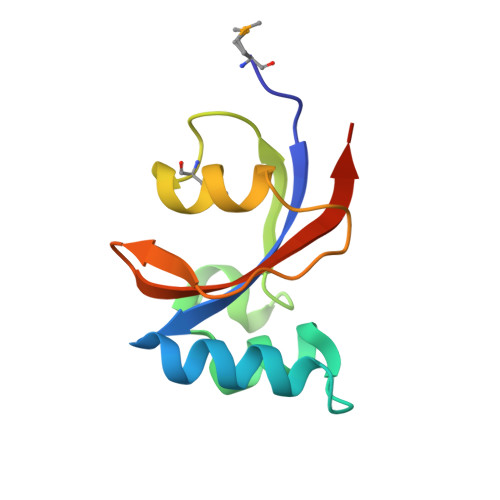
Crystal Structure of Peptidylprolyl Isomerase (PrsA) Fragment from Bacillus anthracis
Minasov, G., Shuvalova, L., Kiryukhina, O., Dubrovska, I., Wiersum, G., Satchell, K.J.F., Center for Structural Genomics of Infectious Diseases (CSGID)To be published.