


The crystal structure of a Beta-lactamase from Escherichia coli CFT073
Tan, K., Wu, R., Endres, M., Joachimiak, A., Center for Structural Genomics of Infectious Diseases (CSGID)To be published.
Experimental Data Snapshot
Entity ID: 1 | |||||
|---|---|---|---|---|---|
| Molecule | Chains | Sequence Length | Organism | Details | Image |
| Beta-lactamase | 361 | Escherichia coli CFT073 | Mutation(s): 0 Gene Names: ampC, c5238 EC: 3.5.2.6 | 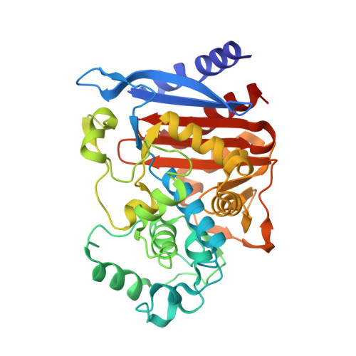 | |
UniProt | |||||
Entity Groups | |||||
| Sequence Clusters | 30% Identity50% Identity70% Identity90% Identity95% Identity100% Identity | ||||
| UniProt Group | P00811 | ||||
Sequence AnnotationsExpand | |||||
Reference Sequence | |||||
| Ligands 3 Unique | |||||
|---|---|---|---|---|---|
| ID | Chains | Name / Formula / InChI Key | 2D Diagram | 3D Interactions | |
| SRT Download:Ideal Coordinates CCD File | L [auth B] | S,R MESO-TARTARIC ACID C4 H6 O6 FEWJPZIEWOKRBE-XIXRPRMCSA-N |  | ||
| SO4 Download:Ideal Coordinates CCD File | G [auth A], H [auth A], M [auth B], N [auth B], O [auth B] | SULFATE ION O4 S QAOWNCQODCNURD-UHFFFAOYSA-L |  | ||
| GOL Download:Ideal Coordinates CCD File | C [auth A] D [auth A] E [auth A] F [auth A] I [auth B] | GLYCEROL C3 H8 O3 PEDCQBHIVMGVHV-UHFFFAOYSA-N |  | ||
| Length ( Å ) | Angle ( ˚ ) |
|---|---|
| a = 128.176 | α = 90 |
| b = 128.176 | β = 90 |
| c = 92.761 | γ = 120 |
| Software Name | Purpose |
|---|---|
| SBC-Collect | data collection |
| PHENIX | refinement |
| HKL-3000 | data reduction |
| HKL-3000 | data scaling |
| HKL-3000 | phasing |
| Funding Organization | Location | Grant Number |
|---|---|---|
| National Institutes of Health/National Institute Of Allergy and Infectious Diseases (NIH/NIAID) | United States | HHSN272201700060C |


