6X9B

Structure of proline utilization A with cis-4-hydroxy-D-proline bound in the L-glutamate-gamma-semialdehyde dehydrogenase active site


Experimental Data Snapshot

  • Method: X-RAY DIFFRACTION
  • Resolution: 1.46 Å
  • R-Value Free: 0.220 
  • R-Value Work: 0.194 
  • R-Value Observed: 0.195 

wwPDB Validation   3D Report Full Report


Ligand Structure Quality Assessment 


This is version 1.2 of the entry. See complete history


Literature

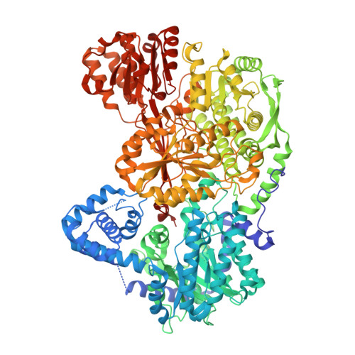
Structural analysis of prolines and hydroxyprolines binding to the l-glutamate-gamma-semialdehyde dehydrogenase active site of bifunctional proline utilization A.

Campbell, A.C.Bogner, A.N.Mao, Y.Becker, D.F.Tanner, J.J.

(2020) Arch Biochem Biophys 698: 108727-108727

  • DOI: https://doi.org/10.1016/j.abb.2020.108727
  • Primary Citation of Related Structures:  
    6X99, 6X9A, 6X9B, 6X9C, 6X9D

  • PubMed Abstract: 

    Proline utilization A (PutA) proteins are bifunctional proline catabolic enzymes that catalyze the 4-electron oxidation of l-proline to l-glutamate using spatially-separated proline dehydrogenase and l-glutamate-γ-semialdehyde dehydrogenase (GSALDH, a.k.a. ALDH4A1) active sites. The observation that l-proline inhibits both the GSALDH activity of PutA and monofunctional GSALDHs motivated us to study the inhibition of PutA by proline stereoisomers and analogs. Here we report five high-resolution crystal structures of PutA with the following ligands bound in the GSALDH active site: d-proline, trans-4-hydroxy-d-proline, cis-4-hydroxy-d-proline, l-proline, and trans-4-hydroxy-l-proline. Three of the structures are of ternary complexes of the enzyme with an inhibitor and either NAD + or NADH. To our knowledge, the NADH complex is the first for any GSALDH. The structures reveal a conserved mode of recognition of the inhibitor carboxylate, which results in the pyrrolidine rings of the d- and l-isomers having different orientations and different hydrogen bonding environments. Activity assays show that the compounds are weak inhibitors with millimolar inhibition constants. Curiously, although the inhibitors occupy the aldehyde binding site, kinetic measurements show the inhibition is uncompetitive. Uncompetitive inhibition may involve proline binding to a remote site or to the enzyme-NADH complex. Together, the structural and kinetic data expand our understanding of how proline-like molecules interact with GSALDH, reveal insight into the relationship between stereochemistry and inhibitor affinity, and demonstrate the pitfalls of inferring the mechanism of inhibition from crystal structures alone.


  • Organizational Affiliation

    Department of Biochemistry, University of Missouri, Columbia, MO, 65211, United States.


Macromolecules
Find similar proteins by:  (by identity cutoff)  |  3D Structure
Entity ID: 1
MoleculeChains Sequence LengthOrganismDetailsImage
Bifunctional protein PutA
A, B
1,235Sinorhizobium meliloti SM11Mutation(s): 0 
Gene Names: putASM11_chr0102
EC: 1.5.5.2 (PDB Primary Data), 1.2.1.88 (PDB Primary Data)
UniProt
Find proteins for F7X6I3 (Sinorhizobium meliloti (strain SM11))
Explore F7X6I3 
Go to UniProtKB:  F7X6I3
Entity Groups  
Sequence Clusters30% Identity50% Identity70% Identity90% Identity95% Identity100% Identity
UniProt GroupF7X6I3
Sequence Annotations
Expand
  • Reference Sequence
Small Molecules
Ligands 7 Unique
IDChains Name / Formula / InChI Key2D Diagram3D Interactions
FAD (Subject of Investigation/LOI)
Query on FAD

Download Ideal Coordinates CCD File 
C [auth A],
I [auth B]
FLAVIN-ADENINE DINUCLEOTIDE
C27 H33 N9 O15 P2
VWWQXMAJTJZDQX-UYBVJOGSSA-N
NAD
Query on NAD

Download Ideal Coordinates CCD File 
E [auth A],
K [auth B]
NICOTINAMIDE-ADENINE-DINUCLEOTIDE
C21 H27 N7 O14 P2
BAWFJGJZGIEFAR-NNYOXOHSSA-N
PGE
Query on PGE

Download Ideal Coordinates CCD File 
J [auth B]TRIETHYLENE GLYCOL
C6 H14 O4
ZIBGPFATKBEMQZ-UHFFFAOYSA-N
UYA (Subject of Investigation/LOI)
Query on UYA

Download Ideal Coordinates CCD File 
H [auth A],
M [auth B]
(4R)-4-hydroxy-D-proline
C5 H9 N O3
PMMYEEVYMWASQN-QWWZWVQMSA-N
PEG
Query on PEG

Download Ideal Coordinates CCD File 
D [auth A]DI(HYDROXYETHYL)ETHER
C4 H10 O3
MTHSVFCYNBDYFN-UHFFFAOYSA-N
SO4
Query on SO4

Download Ideal Coordinates CCD File 
F [auth A],
L [auth B]
SULFATE ION
O4 S
QAOWNCQODCNURD-UHFFFAOYSA-L
MG
Query on MG

Download Ideal Coordinates CCD File 
G [auth A]MAGNESIUM ION
Mg
JLVVSXFLKOJNIY-UHFFFAOYSA-N
Binding Affinity Annotations 
IDSourceBinding Affinity
UYA Binding MOAD:  6X9B Ki: 8.00e+6 (nM) from 1 assay(s)
Experimental Data & Validation

Experimental Data

  • Method: X-RAY DIFFRACTION
  • Resolution: 1.46 Å
  • R-Value Free: 0.220 
  • R-Value Work: 0.194 
  • R-Value Observed: 0.195 
  • Space Group: P 1 21 1
Unit Cell:
Length ( Å )Angle ( ˚ )
a = 100.89α = 90
b = 101.92β = 106.44
c = 126.2γ = 90
Software Package:
Software NamePurpose
Aimlessdata scaling
PHENIXrefinement
PDB_EXTRACTdata extraction
XDSdata reduction
PHENIXphasing

Structure Validation

View Full Validation Report


Ligand Structure Quality Assessment 


Entry History & Funding Information

Deposition Data


Funding OrganizationLocationGrant Number
National Institutes of Health/National Institute of General Medical Sciences (NIH/NIGMS)United StatesR01GM065546

Revision History  (Full details and data files)

  • Version 1.0: 2020-12-30
    Type: Initial release
  • Version 1.1: 2021-01-06
    Changes: Database references
  • Version 1.2: 2023-10-18
    Changes: Data collection, Database references, Refinement description