6X48 | pdb_00006x48

Sortilin-Progranulin Interaction With Compound 17


Experimental Data Snapshot

  • Method: X-RAY DIFFRACTION
  • Resolution: 2.90 Å
  • R-Value Free: 
    0.252 (Depositor), 0.267 (DCC) 
  • R-Value Work: 
    0.193 (Depositor), 0.206 (DCC) 
  • R-Value Observed: 
    0.196 (Depositor) 

Starting Model: experimental
View more details

wwPDB Validation 3D Report Full Report

Validation slider image for 6X48

Ligand Structure Quality Assessment 


This is version 1.2 of the entry. See complete history

Literature

Identification of potent inhibitors of the sortilin-progranulin interaction.

Stachel, S.J.Ginnetti, A.T.Johnson, S.A.Cramer, P.Wang, Y.Bukhtiyarova, M.Krosky, D.Stump, C.Hurzy, D.M.Schlegel, K.A.Cooke, A.J.Allen, S.O'Donnell, G.Ziebell, M.Parthasarathy, G.Getty, K.L.Ho, T.Ou, Y.Jovanovska, A.Carroll, S.S.Pausch, M.Lumb, K.Mosser, S.D.Voleti, B.Klein, D.J.Soisson, S.M.Zerbinatti, C.Coleman, P.J.

(2020) Bioorg Med Chem Lett 30: 127403-127403

  • DOI: https://doi.org/10.1016/j.bmcl.2020.127403
  • Primary Citation Related Structures: 
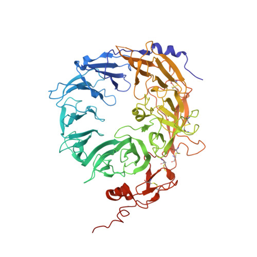
    6X3L, 6X48, 6X4H

  • PubMed Abstract: 

    High-throughput screening methods have been used to identify two novel series of inhibitors that disrupt progranulin binding to sortilin. Exploration of structure-activity relationships (SAR) resulted in compounds with sufficient potency and physicochemical properties to enable co-crystallization with sortilin. These co-crystal structures supported observed SAR trends and provided guidance for additional avenues for designing compounds with additional interactions within the binding site.


  • Organizational Affiliation
    • Department of Medicinal Chemistry, Merck & Co. Inc., PO Box 4, West Point, PA 19486, USA. Electronic address: shawn_stachel@merck.com.

Macromolecule Content 

  • Total Structure Weight: 76.01 kDa 
  • Atom Count: 5,303 
  • Modeled Residue Count: 654 
  • Deposited Residue Count: 663 
  • Unique protein chains: 1

Macromolecules

Find similar proteins by:|  3D Structure
Entity ID: 1
MoleculeChains  Sequence LengthOrganismDetailsImage
Sortilin663Homo sapiensMutation(s): 0 
Gene Names: SORT1
UniProt & NIH Common Fund Data Resources
Find proteins for Q99523 (Homo sapiens)
Explore Q99523 
Go to UniProtKB:  Q99523
PHAROS:  Q99523
GTEx:  ENSG00000134243 
Entity Groups
Sequence Clusters30% Identity50% Identity70% Identity90% Identity95% Identity100% Identity
UniProt GroupQ99523
Glycosylation
Glycosylation Sites: 2Go to GlyGen: Q99523-1
Sequence Annotations
Expand
Reference Sequence

Oligosaccharides

Help  
Entity ID: 2
MoleculeChains Length2D Diagram GlycosylationD Interactions
alpha-D-mannopyranose-(1-6)-beta-D-mannopyranose-(1-4)-2-acetamido-2-deoxy-beta-D-glucopyranose-(1-4)-2-acetamido-2-deoxy-beta-D-glucopyranose
B
4N-Glycosylation
Glycosylation Resources
GlyTouCan: G22573RC
GlyCosmos: G22573RC
GlyGen: G22573RC
Entity ID: 3
MoleculeChains Length2D Diagram GlycosylationD Interactions
beta-D-mannopyranose-(1-4)-2-acetamido-2-deoxy-beta-D-glucopyranose-(1-4)-2-acetamido-2-deoxy-beta-D-glucopyranose
C
3N-Glycosylation
Glycosylation Resources
GlyTouCan: G15407YE
GlyCosmos: G15407YE
GlyGen: G15407YE

Small Molecules

Experimental Data & Validation

Experimental Data

  • Method: X-RAY DIFFRACTION
  • Resolution: 2.90 Å
  • R-Value Free:  0.252 (Depositor), 0.267 (DCC) 
  • R-Value Work:  0.193 (Depositor), 0.206 (DCC) 
  • R-Value Observed: 0.196 (Depositor) 
Space Group: C 1 2 1
Unit Cell:
Length ( Å )Angle ( ˚ )
a = 162.35α = 90
b = 78.49β = 127.23
c = 111.22γ = 90
Software Package:
Software NamePurpose
BUSTERrefinement
PDB_EXTRACTdata extraction
HKL-2000data reduction
HKL-2000data scaling
BUSTERphasing

Structure Validation

View Full Validation Report


Ligand Structure Quality Assessment 


Entry History 

Deposition Data

Revision History  (Full details and data files)

  • Version 1.0: 2020-08-12
    Type: Initial release
  • Version 1.1: 2023-10-18
    Changes: Data collection, Database references, Refinement description
  • Version 1.2: 2024-10-09
    Changes: Structure summary