6X1Q | pdb_00006x1q

1.8 Angstrom resolution structure of b-galactosidase with a 200 kV cryoARM electron microscope


Experimental Data Snapshot

  • Method: ELECTRON MICROSCOPY
  • Resolution: 1.80 Å
  • Aggregation State: PARTICLE 
  • Reconstruction Method: SINGLE PARTICLE 

Starting Model: experimental
View more details

wwPDB Validation 3D Report Full Report

Validation slider image for 6X1Q

This is version 1.2 of the entry. See complete history

Literature

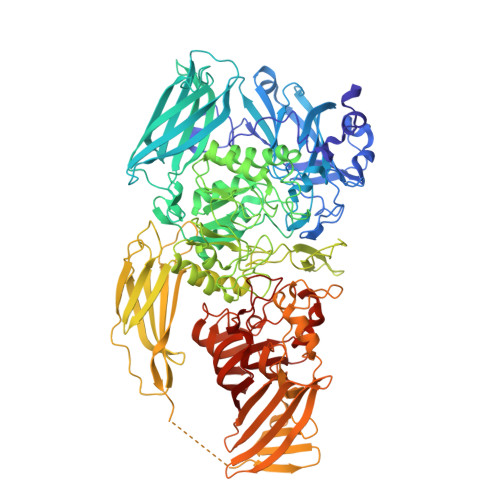
1.8 angstrom resolution structure of beta-galactosidase with a 200 kV CRYO ARM electron microscope.

Merk, A.Fukumura, T.Zhu, X.Darling, J.E.Grisshammer, R.Ognjenovic, J.Subramaniam, S.

(2020) IUCrJ 7: 639-643

  • DOI: https://doi.org/10.1107/S2052252520006855
  • Primary Citation Related Structures: 
    6X1Q

  • PubMed Abstract: 

    We report the determination of the structure of Escherichia coli β-galactosidase at a resolution of ∼1.8 Å using data collected on a 200 kV CRYO ARM microscope equipped with a K3 direct electron detector. The data were collected in a single 24 h session by recording images from an array of 7 × 7 holes at each stage position using the automated data collection program SerialEM . In addition to the expected features such as holes in the densities of aromatic residues, the map also shows density bumps corresponding to the locations of hydrogen atoms. The hydrogen densities are useful in assigning absolute orientations for residues such as glutamine or asparagine by removing the uncertainty in the fitting of the amide groups, and are likely to be especially relevant in the context of structure-guided drug design. These findings validate the use of electron microscopes operating at 200 kV for imaging protein complexes at atomic resolution using cryo-EM.


  • Organizational Affiliation
    • Cancer Research Technology Program, Frederick National Laboratory for Cancer Research, Leidos Biomedical Research, Inc., Frederick, MD 21701, USA.

Macromolecule Content 

  • Total Structure Weight: 465.1 kDa 
  • Atom Count: 35,618 
  • Modeled Residue Count: 4,056 
  • Deposited Residue Count: 4,084 
  • Unique protein chains: 1

Macromolecules

Find similar proteins by:|  3D Structure
Entity ID: 1
MoleculeChains  Sequence LengthOrganismDetailsImage
Beta-galactosidase
A, B, C, D
1,021Escherichia coli K-12Mutation(s): 0 
EC: 3.2.1.23
UniProt
Find proteins for P00722 (Escherichia coli (strain K12))
Explore P00722 
Go to UniProtKB:  P00722
Entity Groups
Sequence Clusters30% Identity50% Identity70% Identity90% Identity95% Identity100% Identity
UniProt GroupP00722
Sequence Annotations
Expand
Reference Sequence

Small Molecules

Ligands 2 Unique
IDChains Name / Formula / InChI Key2D Diagram3D Interactions
MG

Query on MG


Download:Ideal Coordinates CCD File
E [auth A]
F [auth A]
I [auth B]
J [auth B]
M [auth C]
E [auth A],
F [auth A],
I [auth B],
J [auth B],
M [auth C],
N [auth C],
Q [auth D],
R [auth D]
MAGNESIUM ION
Mg
JLVVSXFLKOJNIY-UHFFFAOYSA-N
NA

Query on NA


Download:Ideal Coordinates CCD File
G [auth A]
H [auth A]
K [auth B]
L [auth B]
O [auth C]
G [auth A],
H [auth A],
K [auth B],
L [auth B],
O [auth C],
P [auth C],
S [auth D],
T [auth D]
SODIUM ION
Na
FKNQFGJONOIPTF-UHFFFAOYSA-N

Experimental Data & Validation

Experimental Data

  • Method: ELECTRON MICROSCOPY
  • Resolution: 1.80 Å
  • Aggregation State: PARTICLE 
  • Reconstruction Method: SINGLE PARTICLE 
EM Software:
TaskSoftware PackageVersion
RECONSTRUCTIONRELION3.1

Structure Validation

View Full Validation Report


Entry History 

& Funding Information

Deposition Data


Funding OrganizationLocationGrant Number
National Institutes of Health/National Cancer Institute (NIH/NCI)--

Revision History  (Full details and data files)

  • Version 1.0: 2020-07-01
    Type: Initial release
  • Version 1.1: 2020-08-05
    Changes: Database references, Derived calculations
  • Version 1.2: 2024-03-06
    Changes: Data collection, Database references, Refinement description