6WXL | pdb_00006wxl

Cryo-EM structure of the VRC315 clinical trial, vaccine-elicited, human antibody 1D12 in complex with an H7 SH13 HA trimer


Experimental Data Snapshot

  • Method: ELECTRON MICROSCOPY
  • Resolution: 2.76 Å
  • Aggregation State: PARTICLE 
  • Reconstruction Method: SINGLE PARTICLE 

Starting Models: experimental
View more details

wwPDB Validation 3D Report Full Report

Validation slider image for 6WXL

This is version 1.2 of the entry. See complete history

Literature

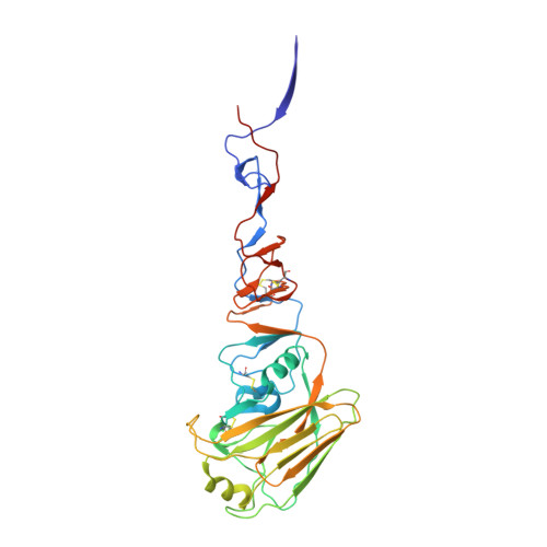
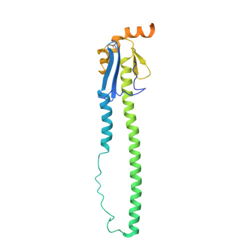
Structure of an influenza group 2-neutralizing antibody targeting the hemagglutinin stem supersite.

Cheung, C.S.Gorman, J.Andrews, S.F.Rawi, R.Reveiz, M.Shen, C.H.Wang, Y.Harris, D.R.Nazzari, A.F.Olia, A.S.Raab, J.Teng, I.T.Verardi, R.Wang, S.Yang, Y.Chuang, G.Y.McDermott, A.B.Zhou, T.Kwong, P.D.

(2022) Structure 

  • DOI: https://doi.org/10.1016/j.str.2022.04.003
  • Primary Citation Related Structures: 
    6WXL

  • PubMed Abstract: 

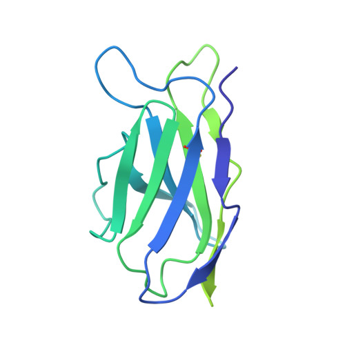
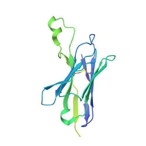
    Several influenza antibodies with broad group 2 neutralization have recently been isolated. Here, we analyze the structure, class, and binding of one of these antibodies from an H7N9 vaccine trial, 315-19-1D12. The cryo-EM structure of 315-19-1D12 Fab in complex with the hemagglutinin (HA) trimer revealed the antibody to recognize the helix A region of the HA stem, at the supersite of vulnerability recognized by group 1-specific and by cross-group-neutralizing antibodies. 315-19-1D12 was derived from HV1-2 and KV2-28 genes and appeared to form a new antibody class. Bioinformatic analysis indicated its group 2 neutralization specificity to be a consequence of four key residue positions. We specifically tested the impact of the group 1-specific N33 glycan, which decreased but did not abolish group 2 binding of 315-19-1D12. Overall, this study highlights the recognition of a broad group 2-neutralizing antibody, revealing unexpected diversity in neutralization specificity for antibodies that recognize the HA stem supersite.


  • Organizational Affiliation
    • Vaccine Research Center, National Institute of Allergy and Infectious Diseases, National Institutes of Health, Bethesda, MD 20892, USA.

Macromolecule Content 

  • Total Structure Weight: 329.85 kDa 
  • Atom Count: 17,016 
  • Modeled Residue Count: 2,175 
  • Deposited Residue Count: 2,985 
  • Unique protein chains: 4

Macromolecules

Find similar proteins by:|  3D Structure
Entity ID: 1
MoleculeChains  Sequence LengthOrganismDetailsImage
Hemagglutinin HA1 chainA,
K [auth C],
L [auth G]
321Influenza A virus (A/Shanghai/JS01/2013(H7N9))Mutation(s): 0 
Gene Names: HA
UniProt
Find proteins for A0A067Y6L0 (Influenza A virus)
Explore A0A067Y6L0 
Go to UniProtKB:  A0A067Y6L0
Entity Groups
Sequence Clusters30% Identity50% Identity70% Identity90% Identity95% Identity100% Identity
UniProt GroupA0A067Y6L0
Glycosylation
Glycosylation Sites: 1
Sequence Annotations
Expand
Reference Sequence
Find similar proteins by:|  3D Structure
Entity ID: 2
MoleculeChains  Sequence LengthOrganismDetailsImage
Hemagglutinin HA2 chainB,
E [auth D],
H [auth I]
221Influenza A virus (A/Shanghai/JS01/2013(H7N9))Mutation(s): 0 
Gene Names: HA
UniProt
Find proteins for A0A067Y6L0 (Influenza A virus)
Explore A0A067Y6L0 
Go to UniProtKB:  A0A067Y6L0
Entity Groups
Sequence Clusters30% Identity50% Identity70% Identity90% Identity95% Identity100% Identity
UniProt GroupA0A067Y6L0
Glycosylation
Glycosylation Sites: 2
Sequence Annotations
Expand
Reference Sequence
Find similar proteins by:|  3D Structure
Entity ID: 3
MoleculeChains  Sequence LengthOrganismDetailsImage
1D12 Light chainC [auth L],
F [auth E],
I [auth J]
221Homo sapiensMutation(s): 0 
Find similar proteins by:|  3D Structure
Entity ID: 4
MoleculeChains  Sequence LengthOrganismDetailsImage
1D21 Heavy chainD [auth H],
G [auth F],
J [auth K]
232Homo sapiensMutation(s): 0 

Small Molecules

Ligands 1 Unique
IDChains Name / Formula / InChI Key2D Diagram3D Interactions
NAG

Query on NAG


Download:Ideal Coordinates CCD File
M [auth A]
N [auth B]
O [auth B]
P [auth D]
Q [auth D]
M [auth A],
N [auth B],
O [auth B],
P [auth D],
Q [auth D],
R [auth I],
S [auth I],
T [auth C],
U [auth G]
2-acetamido-2-deoxy-beta-D-glucopyranose
C8 H15 N O6
OVRNDRQMDRJTHS-FMDGEEDCSA-N

Experimental Data & Validation

Experimental Data

  • Method: ELECTRON MICROSCOPY
  • Resolution: 2.76 Å
  • Aggregation State: PARTICLE 
  • Reconstruction Method: SINGLE PARTICLE 
EM Software:
TaskSoftware PackageVersion
RECONSTRUCTIONcryoSPARC2.12
MODEL REFINEMENTPHENIX

Structure Validation

View Full Validation Report


Entry History 

& Funding Information

Deposition Data


Funding OrganizationLocationGrant Number
National Institutes of Health/National Institute of General Medical Sciences (NIH/NIGMS)United StatesGM103310

Revision History  (Full details and data files)

  • Version 1.0: 2021-06-09
    Type: Initial release
  • Version 1.1: 2022-06-22
    Changes: Database references
  • Version 1.2: 2024-11-20
    Changes: Data collection, Refinement description, Structure summary