6WVT | pdb_00006wvt

Structural basis of alphaE-catenin - F-actin catch bond behavior


Experimental Data Snapshot

  • Method: ELECTRON MICROSCOPY
  • Resolution: 3.56 Å
  • Aggregation State: HELICAL ARRAY 
  • Reconstruction Method: HELICAL 

Starting Models: experimental
View more details

wwPDB Validation 3D Report Full Report

Validation slider image for 6WVT

This is version 1.1 of the entry. See complete history

Literature

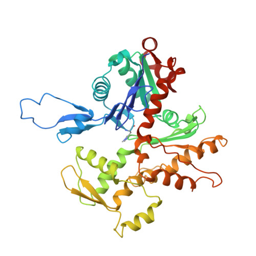
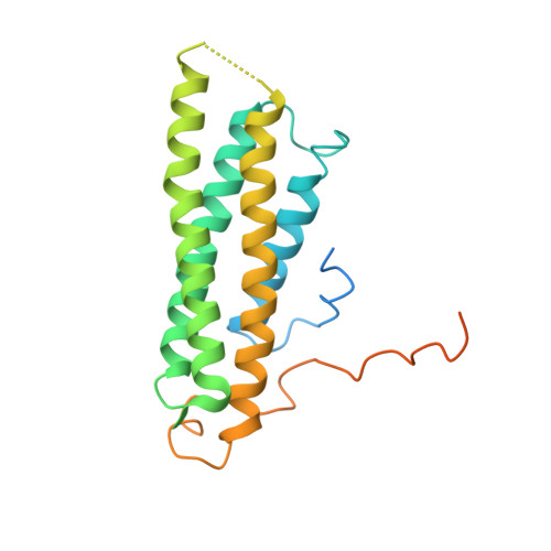
Structural basis of alpha E-catenin-F-actin catch bond behavior.

Xu, X.P.Pokutta, S.Torres, M.Swift, M.F.Hanein, D.Volkmann, N.Weis, W.I.

(2020) Elife 9

  • DOI: https://doi.org/10.7554/eLife.60878
  • Primary Citation Related Structures: 
    6WVT

  • PubMed Abstract: 

    Cell-cell and cell-matrix junctions transmit mechanical forces during tissue morphogenesis and homeostasis. α-Catenin links cell-cell adhesion complexes to the actin cytoskeleton, and mechanical load strengthens its binding to F-actin in a direction-sensitive manner. Specifically, optical trap experiments revealed that force promotes a transition between weak and strong actin-bound states. Here, we describe the cryo-electron microscopy structure of the F-actin-bound αE-catenin actin-binding domain, which in solution forms a five-helix bundle. In the actin-bound structure, the first helix of the bundle dissociates and the remaining four helices and connecting loops rearrange to form the interface with actin. Deletion of the first helix produces strong actin binding in the absence of force, suggesting that the actin-bound structure corresponds to the strong state. Our analysis explains how mechanical force applied to αE-catenin or its homolog vinculin favors the strongly bound state, and the dependence of catch bond strength on the direction of applied force.


  • Organizational Affiliation
    • Scintillon Institute, San Diego, United States.

Macromolecule Content 

  • Total Structure Weight: 411.36 kDa 
  • Atom Count: 25,248 
  • Modeled Residue Count: 3,240 
  • Deposited Residue Count: 3,678 
  • Unique protein chains: 2

Macromolecules

Find similar proteins by:|  3D Structure
Entity ID: 1
MoleculeChains  Sequence LengthOrganismDetailsImage
Actin, alpha skeletal muscle377Oryctolagus cuniculusMutation(s): 0 
EC: 3.6.4
UniProt
Find proteins for P68135 (Oryctolagus cuniculus)
Explore P68135 
Go to UniProtKB:  P68135
Entity Groups
Sequence Clusters30% Identity50% Identity70% Identity90% Identity95% Identity100% Identity
UniProt GroupP68135
Sequence Annotations
Expand
Reference Sequence
Find similar proteins by:|  3D Structure
Entity ID: 2
MoleculeChains  Sequence LengthOrganismDetailsImage
Catenin alpha-1236Mus musculusMutation(s): 0 
Gene Names: Ctnna1Catna1
UniProt
Find proteins for P26231 (Mus musculus)
Explore P26231 
Go to UniProtKB:  P26231
Entity Groups
Sequence Clusters30% Identity50% Identity70% Identity90% Identity95% Identity100% Identity
UniProt GroupP26231
Sequence Annotations
Expand
Reference Sequence

Small Molecules

Ligands 2 Unique
IDChains Name / Formula / InChI Key2D Diagram3D Interactions
ADP

Query on ADP


Download:Ideal Coordinates CCD File
N [auth B]
P [auth D]
R [auth E]
T [auth F]
V [auth H]
N [auth B],
P [auth D],
R [auth E],
T [auth F],
V [auth H],
X [auth I]
ADENOSINE-5'-DIPHOSPHATE
C10 H15 N5 O10 P2
XTWYTFMLZFPYCI-KQYNXXCUSA-N
MG

Query on MG


Download:Ideal Coordinates CCD File
M [auth B]
O [auth D]
Q [auth E]
S [auth F]
U [auth H]
M [auth B],
O [auth D],
Q [auth E],
S [auth F],
U [auth H],
W [auth I]
MAGNESIUM ION
Mg
JLVVSXFLKOJNIY-UHFFFAOYSA-N
Modified Residues  1 Unique
IDChains TypeFormula2D DiagramParent
HIC
Query on HIC
A [auth B]
B [auth D]
C [auth E]
D [auth F]
E [auth H]
A [auth B],
B [auth D],
C [auth E],
D [auth F],
E [auth H],
F [auth I]
L-PEPTIDE LINKINGC7 H11 N3 O2HIS

Experimental Data & Validation

Experimental Data

  • Method: ELECTRON MICROSCOPY
  • Resolution: 3.56 Å
  • Aggregation State: HELICAL ARRAY 
  • Reconstruction Method: HELICAL 
EM Software:
TaskSoftware PackageVersion
MODEL REFINEMENTPHENIX
RECONSTRUCTIONRELION3

Structure Validation

View Full Validation Report


Entry History 

& Funding Information

Deposition Data


Funding OrganizationLocationGrant Number
National Institutes of Health/National Institute of General Medical Sciences (NIH/NIGMS)United StatesGM118326

Revision History  (Full details and data files)

  • Version 1.0: 2020-10-07
    Type: Initial release
  • Version 1.1: 2025-04-02
    Changes: Data collection, Database references, Refinement description, Structure summary