6WUE

Tetragonal crystal form of SbtB from Synechocystis PCC6803


Experimental Data Snapshot

  • Method: X-RAY DIFFRACTION
  • Resolution: 1.80 Å
  • R-Value Free: 0.205 
  • R-Value Work: 0.186 
  • R-Value Observed: 0.188 

wwPDB Validation   3D Report Full Report


Ligand Structure Quality Assessment 


This is version 1.2 of the entry. See complete history


Literature

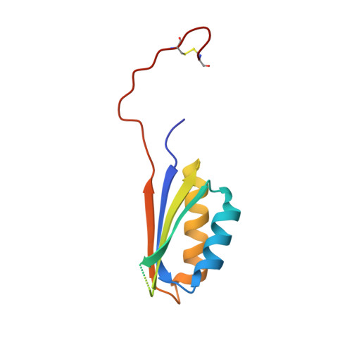
Tetragonal crystal form of the cyanobacterial bicarbonate-transporter regulator SbtB from Synechocystis sp. PCC 6803.

Bu, G.Simmons, C.R.Nielsen, D.R.Nannenga, B.L.

(2020) Acta Crystallogr F Struct Biol Commun 76: 438-443

  • DOI: https://doi.org/10.1107/S2053230X20010523
  • Primary Citation of Related Structures:  
    6WUE

  • PubMed Abstract: 

    The P II -like protein SbtB has been identified as a regulator of SbtA, which is one of the key bicarbonate transporters in cyanobacteria. While SbtB from Synechocystis sp. PCC 6803 has previously been shown to be a trimer, a new crystal form is reported here which crystallizes in what is thought to be a non-native tetramer in the crystal, with the C-terminus in an extended conformation. The crystal structure shows the formation of an intermolecular disulfide bond at Cys94 between SbtB monomers, which may stabilize this conformation in the crystal. This motivates the need for future studies to investigate the potential role that the oxidation and reduction of these cysteines may play in the activation and/or function of SbtB.


  • Organizational Affiliation

    Chemical Engineering, School for Engineering of Matter, Transport and Energy, Arizona State University, Tempe, AZ 85287, USA.


Macromolecules
Find similar proteins by:  (by identity cutoff)  |  3D Structure
Entity ID: 1
MoleculeChains Sequence LengthOrganismDetailsImage
Membrane-associated protein slr1513
A, B
110Synechocystis sp. PCC 6803Mutation(s): 0 
Gene Names: slr1513
UniProt
Find proteins for P73954 (Synechocystis sp. (strain PCC 6803 / Kazusa))
Explore P73954 
Go to UniProtKB:  P73954
Entity Groups  
Sequence Clusters30% Identity50% Identity70% Identity90% Identity95% Identity100% Identity
UniProt GroupP73954
Sequence Annotations
Expand
  • Reference Sequence
Experimental Data & Validation

Experimental Data

  • Method: X-RAY DIFFRACTION
  • Resolution: 1.80 Å
  • R-Value Free: 0.205 
  • R-Value Work: 0.186 
  • R-Value Observed: 0.188 
  • Space Group: P 43 21 2
Unit Cell:
Length ( Å )Angle ( ˚ )
a = 51.571α = 90
b = 51.571β = 90
c = 178.412γ = 90
Software Package:
Software NamePurpose
PHENIXrefinement
HKL-2000data reduction
HKL-2000data scaling
PHASERphasing

Structure Validation

View Full Validation Report


Ligand Structure Quality Assessment 


Entry History 

Deposition Data

Revision History  (Full details and data files)

  • Version 1.0: 2020-08-26
    Type: Initial release
  • Version 1.1: 2020-09-16
    Changes: Database references
  • Version 1.2: 2023-10-18
    Changes: Data collection, Database references, Refinement description