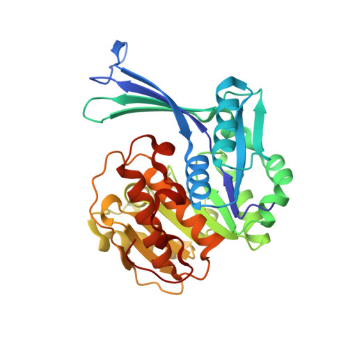
Conformational flexibility of human ribokinase captured in seven crystal structures.
Akanmori, N.N., Junop, M.S., Gupta, R.S., Park, J.(2025) Int J Biol Macromol 299: 140109-140109
- PubMed: 39837438 Search on PubMed
- DOI: https://doi.org/10.1016/j.ijbiomac.2025.140109
- Primary Citation Related Structures:
5BYC, 5BYD, 5BYE, 5C3Z, 5C40, 5C41, 6WK0 - PubMed Abstract:
d-ribose is a critical sugar substrate involved in the biosynthesis of nucleotides, amino acids, and cofactors, with its phosphorylation to ribose-5-phosphate by ribokinase (RK) constituting the initial step in its metabolism. RK is conserved across all domains of life, and its activity is significantly enhanced by monovalent metal (M + ) ions, particularly K + , although the precise mechanism of this activation remains unclear. In this study, we present several crystal structures of human RK in both unliganded and substrate-bound states, offering detailed insights into its substrate binding process, reaction mechanism, and conformational changes throughout the catalytic cycle. Notably, bound ATP exhibited significant conformational flexibility in its triphosphate moiety, a feature shared with other RK homologues, suggesting that achieving a catalytically productive triphosphate configuration plays a key role in regulating enzyme activity. We also identified a unique conformational change in the M + ion binding loop of human RK, specifically the flipping of the Gly306-Thr307 peptide plane, likely influenced by the ionic radius of the bound ion. These findings provide new insights into the RK reaction mechanism and its activation by M + ions, paving the way for future investigations into the allosteric regulation of human RK and related sugar kinase enzymes.
- Department of Biochemistry, Memorial University of Newfoundland, 45 Arctic Avenue, St. John's, Newfoundland and Labrador, Canada.
Organizational Affiliation: