


Crystal structures of human ribokinase
Park, J.To be published.
Experimental Data Snapshot
Starting Model: experimental
View more details
Entity ID: 1 | |||||
|---|---|---|---|---|---|
| Molecule | Chains | Sequence Length | Organism | Details | Image |
| Ribokinase | 317 | Homo sapiens | Mutation(s): 0 Gene Names: RBKS, RBSK EC: 2.7.1.15 | 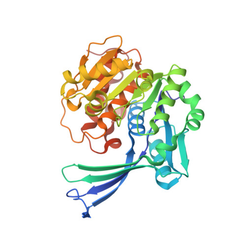 | |
UniProt & NIH Common Fund Data Resources | |||||
PHAROS: Q9H477 GTEx: ENSG00000171174 | |||||
Entity Groups | |||||
| Sequence Clusters | 30% Identity50% Identity70% Identity90% Identity95% Identity100% Identity | ||||
| UniProt Group | Q9H477 | ||||
Sequence AnnotationsExpand | |||||
Reference Sequence | |||||
| Ligands 4 Unique | |||||
|---|---|---|---|---|---|
| ID | Chains | Name / Formula / InChI Key | 2D Diagram | 3D Interactions | |
| A12 (Subject of Investigation/LOI) Download:Ideal Coordinates CCD File | C [auth A], K [auth B] | PHOSPHOMETHYLPHOSPHONIC ACID ADENOSYL ESTER C11 H17 N5 O9 P2 OLCWZBFDIYXLAA-IOSLPCCCSA-N |  | ||
| GOL Download:Ideal Coordinates CCD File | D [auth A] E [auth A] F [auth A] G [auth A] H [auth A] | GLYCEROL C3 H8 O3 PEDCQBHIVMGVHV-UHFFFAOYSA-N |  | ||
| MG (Subject of Investigation/LOI) Download:Ideal Coordinates CCD File | J [auth A], N [auth B] | MAGNESIUM ION Mg JLVVSXFLKOJNIY-UHFFFAOYSA-N |  | ||
| NA (Subject of Investigation/LOI) Download:Ideal Coordinates CCD File | I [auth A], M [auth B] | SODIUM ION Na FKNQFGJONOIPTF-UHFFFAOYSA-N |  | ||
| Length ( Å ) | Angle ( ˚ ) |
|---|---|
| a = 46.11 | α = 90 |
| b = 71.86 | β = 91.78 |
| c = 91.43 | γ = 90 |
| Software Name | Purpose |
|---|---|
| REFMAC | refinement |
| XDS | data reduction |
| XSCALE | data scaling |
| PHASER | phasing |


