6WJP | pdb_00006wjp

Crystal structure of Arginine Repressor P115Q mutant from the pathogenic bacterium Corynebacterium pseudotuberculosis bound to arginine


Experimental Data Snapshot

  • Method: X-RAY DIFFRACTION
  • Resolution: 1.70 Å
  • R-Value Free: 
    0.197 (Depositor), 0.203 (DCC) 
  • R-Value Work: 
    0.173 (Depositor), 0.184 (DCC) 
  • R-Value Observed: 
    0.180 (Depositor) 

Starting Model: experimental
View more details

wwPDB Validation 3D Report Full Report

Validation slider image for 6WJP

Ligand Structure Quality Assessment 


This is version 1.1 of the entry. See complete history

Literature

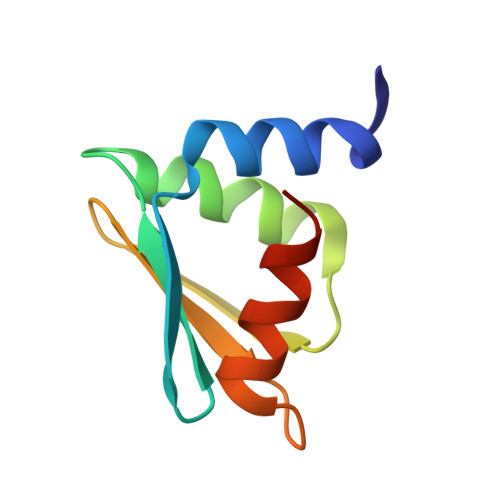
A single P115Q mutation modulates specificity in the Corynebacterium pseudotuberculosis arginine repressor.

Mariutti, R.B.Hernandez-Gonzalez, J.E.Nascimento, A.F.Z.de Morais, M.A.B.Murakami, M.T.Carareto, C.M.A.Arni, R.K.

(2020) Biochim Biophys Acta Gen Subj 1864: 129597

  • DOI: https://doi.org/10.1016/j.bbagen.2020.129597
  • Primary Citation Related Structures: 
    6WJO, 6WJP

  • PubMed Abstract: 

    The arginine repressor (ArgR) regulates the expression of genes involved in arginine biosynthesis. Upon attaining a threshold concentration of arginine in the cytoplasm, the trimeric C-terminal domain of ArgR binds three arginines in a shallow surface cleft and subsequently hexamerizes forming a dimer of trimers containing six Arg co-repressor molecules which are buried at the subunit interfaces. The N-terminal domains of this complex bind to the DNA promoter thereby interrupting the transcription of the genes related to Arg biosynthesis. The crystal structures of the wild type and mutant Pro115Gln ArgR from Corynebacterium pseudotuberculosis determined at 1.7 Å demonstrate that a single amino acid substitution switches co-repressor specificity from Tyr to Arg. Molecular dynamics simulations indicate that the first step, i.e., the binding of the co-repressor, occurs in the trimeric state and that Pro115Gln ArgR preferentially binds Arg. It was also shown that, in Pro115 ArgR hexamers, the concomitant binding of sodium ions shifts selectivity to Tyr. Structural data combined with phylogenetic analyses of ArgR from C. pseudotuberculosis suggest that substitutions in the binding pocket at position 115 may alter its specificity for amino acids and that the length of the protein interdomain linker can provide further functional flexibility. These results support the existence of alternative ArgR regulatory mechanisms in this pathogenic bacterium.


  • Organizational Affiliation
    • Multiuser Center for Biomolecular Innovation, IBILCE/UNESP, São José do Rio Preto, SP, Brazil. Electronic address: ricardomariutti@yahoo.com.br.

Macromolecule Content 

  • Total Structure Weight: 18.56 kDa 
  • Atom Count: 1,308 
  • Modeled Residue Count: 159 
  • Deposited Residue Count: 166 
  • Unique protein chains: 1

Macromolecules

Find similar proteins by:|  3D Structure
Entity ID: 1
MoleculeChains  Sequence LengthOrganismDetailsImage
Arginine repressor
A, B
83Corynebacterium pseudotuberculosis C231Mutation(s): 1 
Gene Names: argRCpC231_0953
UniProt
Find proteins for D9QA55 (Corynebacterium pseudotuberculosis (strain C231))
Explore D9QA55 
Go to UniProtKB:  D9QA55
Entity Groups
Sequence Clusters30% Identity50% Identity70% Identity90% Identity95% Identity100% Identity
UniProt GroupD9QA55
Sequence Annotations
Expand
Reference Sequence

Small Molecules

Ligands 4 Unique
IDChains Name / Formula / InChI Key2D Diagram3D Interactions
ARG
(Subject of Investigation/LOI)

Query on ARG


Download:Ideal Coordinates CCD File
C [auth A],
E [auth B]
ARGININE
C6 H15 N4 O2
ODKSFYDXXFIFQN-BYPYZUCNSA-O
TRS

Query on TRS


Download:Ideal Coordinates CCD File
G [auth B]2-AMINO-2-HYDROXYMETHYL-PROPANE-1,3-DIOL
C4 H12 N O3
LENZDBCJOHFCAS-UHFFFAOYSA-O
GOL

Query on GOL


Download:Ideal Coordinates CCD File
F [auth B]GLYCEROL
C3 H8 O3
PEDCQBHIVMGVHV-UHFFFAOYSA-N
ACT

Query on ACT


Download:Ideal Coordinates CCD File
D [auth A]ACETATE ION
C2 H3 O2
QTBSBXVTEAMEQO-UHFFFAOYSA-M

Experimental Data & Validation

Experimental Data

  • Method: X-RAY DIFFRACTION
  • Resolution: 1.70 Å
  • R-Value Free:  0.197 (Depositor), 0.203 (DCC) 
  • R-Value Work:  0.173 (Depositor), 0.184 (DCC) 
  • R-Value Observed: 0.180 (Depositor) 
Space Group: P 21 3
Unit Cell:
Length ( Å )Angle ( ˚ )
a = 83.341α = 90
b = 83.341β = 90
c = 83.341γ = 90
Software Package:
Software NamePurpose
XDSdata reduction
XSCALEdata scaling
PHASERphasing
PHENIXrefinement
PDB_EXTRACTdata extraction

Structure Validation

View Full Validation Report


Ligand Structure Quality Assessment 


Entry History 

& Funding Information

Deposition Data


Funding OrganizationLocationGrant Number
Brazilian National Council for Scientific and Technological Development (CNPq)Brazil307338/2014-2
Brazilian National Council for Scientific and Technological Development (CNPq)Brazil309940/2019-2
Brazilian National Council for Scientific and Technological Development (CNPq)Brazil303455/2017-9
Sao Paulo Research Foundation (FAPESP)Brazil2016/19995-0
Sao Paulo Research Foundation (FAPESP)Brazil2018/10736-8
Sao Paulo Research Foundation (FAPESP)Brazil2018/07977-3
Sao Paulo Research Foundation (FAPESP)Brazil2015/13765-0
Sao Paulo Research Foundation (FAPESP)Brazil2015/18868-2
Sao Paulo Research Foundation (FAPESP)Brazil2016/24587-9

Revision History  (Full details and data files)

  • Version 1.0: 2020-04-22
    Type: Initial release
  • Version 1.1: 2023-10-18
    Changes: Advisory, Data collection, Database references, Refinement description