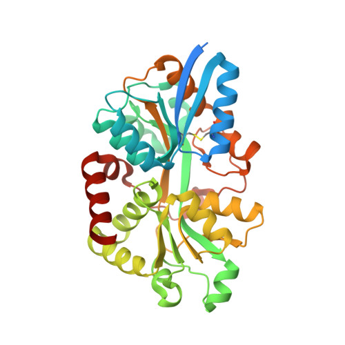
6WCE

Structure of the periplasmic binding protein P5PA


Experimental Data Snapshot

  • Method: X-RAY DIFFRACTION
  • Resolution: 1.75 Å
  • R-Value Free: 0.195 
  • R-Value Work: 0.161 
  • R-Value Observed: 0.162 

Starting Model: experimental
View more details

wwPDB Validation   3D Report Full Report


Ligand Structure Quality Assessment 


This is version 1.2 of the entry. See complete history


Literature

Actinobacillus utilizes a binding protein-dependent ABC transporter to acquire the active form of vitamin B 6 .

Pan, C.Zimmer, A.Shah, M.Huynh, M.S.Lai, C.C.Sit, B.Hooda, Y.Curran, D.M.Moraes, T.F.

(2021) J Biol Chem 297: 101046-101046

  • DOI: https://doi.org/10.1016/j.jbc.2021.101046
  • Primary Citation of Related Structures:  
    6WCE

  • PubMed Abstract: 

    Bacteria require high-efficiency uptake systems to survive and proliferate in nutrient-limiting environments, such as those found in host organisms. ABC transporters in the bacterial plasma membrane provide a mechanism for transport of many substrates. In this study, we examine an operon containing a periplasmic binding protein in Actinobacillus for its potential role in nutrient acquisition. The electron density map of 1.76 Å resolution obtained from the crystal structure of the periplasmic binding protein was best fit with a molecular model containing a pyridoxal-5'-phosphate (P5P/pyridoxal phosphate/the active form of vitamin B 6 ) ligand within the protein's binding site. The identity of the P5P bound to this periplasmic binding protein was verified by isothermal titration calorimetry, microscale thermophoresis, and mass spectrometry, leading us to name the protein P5PA and the operon P5PAB. To illustrate the functional utility of this uptake system, we introduced the P5PAB operon from Actinobacillus pleuropneumoniae into an Escherichia coli K-12 strain that was devoid of a key enzyme required for P5P synthesis. The growth of this strain at low levels of P5P supports the functional role of this operon in P5P uptake. This is the first report of a dedicated P5P bacterial uptake system, but through bioinformatics, we discovered homologs mainly within pathogenic representatives of the Pasteurellaceae family, suggesting that this operon exists more widely outside the Actinobacillus genus.


  • Organizational Affiliation

    Department of Biochemistry, University of Toronto, Toronto, Ontario, Canada.


Macromolecules
Find similar proteins by:  (by identity cutoff)  |  3D Structure
Entity ID: 1
MoleculeChains Sequence LengthOrganismDetailsImage
Pyridoxal-5-phosphate binding protein A (P5PA)355Actinobacillus pleuropneumoniaeMutation(s): 0 
UniProt
Find proteins for B0BUC2 (Actinobacillus pleuropneumoniae serotype 3 (strain JL03))
Explore B0BUC2 
Go to UniProtKB:  B0BUC2
Entity Groups  
Sequence Clusters30% Identity50% Identity70% Identity90% Identity95% Identity100% Identity
UniProt GroupB0BUC2
Sequence Annotations
Expand
  • Reference Sequence
Small Molecules
Ligands 1 Unique
IDChains Name / Formula / InChI Key2D Diagram3D Interactions
PLP (Subject of Investigation/LOI)
Query on PLP

Download Ideal Coordinates CCD File 
B [auth A]PYRIDOXAL-5'-PHOSPHATE
C8 H10 N O6 P
NGVDGCNFYWLIFO-UHFFFAOYSA-N
Experimental Data & Validation

Experimental Data

  • Method: X-RAY DIFFRACTION
  • Resolution: 1.75 Å
  • R-Value Free: 0.195 
  • R-Value Work: 0.161 
  • R-Value Observed: 0.162 
  • Space Group: P 1 21 1
Unit Cell:
Length ( Å )Angle ( ˚ )
a = 51.468α = 90
b = 42.273β = 106.818
c = 69.601γ = 90
Software Package:
Software NamePurpose
PHENIXrefinement
XSCALEdata scaling
PDB_EXTRACTdata extraction
iMOSFLMdata reduction
PHASERphasing

Structure Validation

View Full Validation Report


Ligand Structure Quality Assessment 


Entry History & Funding Information

Deposition Data


Funding OrganizationLocationGrant Number
Natural Sciences and Engineering Research Council (NSERC, Canada)CanadaRGPIN-2018-06546

Revision History  (Full details and data files)

  • Version 1.0: 2021-08-25
    Type: Initial release
  • Version 1.1: 2021-09-22
    Changes: Database references
  • Version 1.2: 2023-10-18
    Changes: Data collection, Refinement description