6W9G | pdb_00006w9g

Crystal Structure of the Fab fragment of humanized 5c8 antibody containing the fluorescent non-canonical amino acid L-(7-hydroxycoumarin-4-yl)ethylglycine in complex with CD40L at pH 6.8


Experimental Data Snapshot

  • Method: X-RAY DIFFRACTION
  • Resolution: 1.82 Å
  • R-Value Free: 
    0.207 (Depositor), 0.211 (DCC) 
  • R-Value Work: 
    0.179 (Depositor), 0.187 (DCC) 
  • R-Value Observed: 
    0.180 (Depositor) 

Starting Model: experimental
View more details

wwPDB Validation 3D Report Full Report

Validation slider image for 6W9G

This is version 1.3 of the entry. See complete history

Literature

Structural Insights into How Protein Environments Tune the Spectroscopic Properties of a Noncanonical Amino Acid Fluorophore.

Henderson, J.N.Simmons, C.R.Fahmi, N.E.Jeffs, J.W.Borges, C.R.Mills, J.H.

(2020) Biochemistry 59: 3401-3410

  • DOI: https://doi.org/10.1021/acs.biochem.0c00474
  • Primary Citation Related Structures: 
    6BJZ, 6W4W, 6W5A, 6W9G

  • PubMed Abstract: 

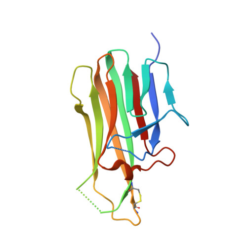
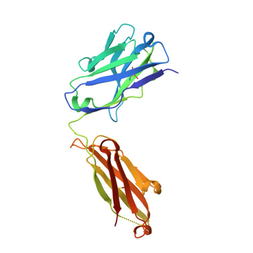
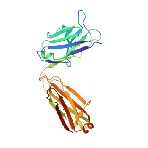
    Genetically encoded fluorescent noncanonical amino acids (fNCAAs) could be used to develop novel fluorescent sensors of protein function. Previous efforts toward this goal have been limited by the lack of extensive physicochemical and structural characterizations of protein-based sensors containing fNCAAs. Here, we report the steady-state spectroscopic properties and first structural analyses of an fNCAA-containing Fab fragment of the 5c8 antibody, which binds human CD40L. A previously reported 5c8 variant in which the light chain residue Ile L 98 is replaced with the fNCAA l-(7-hydroxycoumarin-4-yl)ethylglycine (7-HCAA) exhibits a 1.7-fold increase in fluorescence upon antigen binding. Determination and comparison of the apparent p K a s of 7-HCAA in the unbound and bound forms indicate that the observed increase in fluorescence is not the result of perturbations in p K a . Crystal structures of the fNCAA-containing Fab in the apo and bound forms reveal interactions between the 7-HCAA side chain and surrounding residues that are disrupted upon antigen binding. This structural characterization not only provides insight into the manner in which protein environments can modulate the fluorescence properties of 7-HCAA but also could serve as a starting point for the rational design of new fluorescent protein-based reporters of protein function.


  • Organizational Affiliation
    • The Biodesign Center for Molecular Design and Biomimetics, Arizona State University, Tempe, Arizona 85287, United States.

Macromolecule Content 

  • Total Structure Weight: 198.47 kDa 
  • Atom Count: 14,802 
  • Modeled Residue Count: 1,696 
  • Deposited Residue Count: 1,770 
  • Unique protein chains: 3

Macromolecules

Find similar proteins by:|  3D Structure
Entity ID: 1
MoleculeChains  Sequence LengthOrganismDetailsImage
CD40 ligand
A, B, C
146Homo sapiensMutation(s): 0 
Gene Names: CD40LGCD40LTNFSF5TRAP
UniProt & NIH Common Fund Data Resources
Find proteins for P29965 (Homo sapiens)
Explore P29965 
Go to UniProtKB:  P29965
PHAROS:  P29965
GTEx:  ENSG00000102245 
Entity Groups
Sequence Clusters30% Identity50% Identity70% Identity90% Identity95% Identity100% Identity
UniProt GroupP29965
Glycosylation
Glycosylation Sites: 1Go to GlyGen: P29965-1
Sequence Annotations
Expand
Reference Sequence
Find similar proteins by:|  3D Structure
Entity ID: 2
MoleculeChains  Sequence LengthOrganismDetailsImage
5c8* Fab (heavy chain)D [auth H],
F [auth K],
H [auth X]
226Homo sapiensMutation(s): 0 
Find similar proteins by:|  3D Structure
Entity ID: 3
MoleculeChains  Sequence LengthOrganismDetailsImage
5c8* Fab (light chain)E [auth L],
G [auth M],
I [auth Y]
218Homo sapiensMutation(s): 0 

Oligosaccharides

Help  
Entity ID: 4
MoleculeChains Length2D Diagram GlycosylationD Interactions
alpha-D-mannopyranose-(1-2)-alpha-D-mannopyranose-(1-6)-alpha-D-mannopyranose-(1-6)-beta-D-mannopyranose-(1-4)-2-acetamido-2-deoxy-beta-D-glucopyranose-(1-4)-2-acetamido-2-deoxy-beta-D-glucopyranoseJ [auth D],
K [auth E],
L [auth F]
6N-Glycosylation
Glycosylation Resources
GlyTouCan: G02763QD
GlyCosmos: G02763QD
GlyGen: G02763QD

Small Molecules

Ligands 5 Unique
IDChains Name / Formula / InChI Key2D Diagram3D Interactions
1PE

Query on 1PE


Download:Ideal Coordinates CCD File
X [auth C]PENTAETHYLENE GLYCOL
C10 H22 O6
JLFNLZLINWHATN-UHFFFAOYSA-N
ETE

Query on ETE


Download:Ideal Coordinates CCD File
NA [auth M],
Q [auth A],
T [auth B]
2-{2-[2-2-(METHOXY-ETHOXY)-ETHOXY]-ETHOXY}-ETHANOL
C9 H20 O5
ZNYRFEPBTVGZDN-UHFFFAOYSA-N
TRS

Query on TRS


Download:Ideal Coordinates CCD File
N [auth A],
O [auth A]
2-AMINO-2-HYDROXYMETHYL-PROPANE-1,3-DIOL
C4 H12 N O3
LENZDBCJOHFCAS-UHFFFAOYSA-O
PEG

Query on PEG


Download:Ideal Coordinates CCD File
AA [auth L]
M [auth A]
QA [auth Y]
R [auth B]
RA [auth Y]
AA [auth L],
M [auth A],
QA [auth Y],
R [auth B],
RA [auth Y],
SA [auth Y],
U [auth C],
V [auth C]
DI(HYDROXYETHYL)ETHER
C4 H10 O3
MTHSVFCYNBDYFN-UHFFFAOYSA-N
TMO

Query on TMO


Download:Ideal Coordinates CCD File
BA [auth L]
CA [auth L]
DA [auth L]
EA [auth L]
FA [auth K]
BA [auth L],
CA [auth L],
DA [auth L],
EA [auth L],
FA [auth K],
GA [auth K],
HA [auth K],
IA [auth K],
JA [auth M],
KA [auth M],
LA [auth M],
MA [auth M],
OA [auth X],
P [auth A],
PA [auth X],
S [auth B],
TA [auth Y],
UA [auth Y],
W [auth C],
Y [auth H],
Z [auth H]
trimethylamine oxide
C3 H9 N O
UYPYRKYUKCHHIB-UHFFFAOYSA-N

Experimental Data & Validation

Experimental Data

  • Method: X-RAY DIFFRACTION
  • Resolution: 1.82 Å
  • R-Value Free:  0.207 (Depositor), 0.211 (DCC) 
  • R-Value Work:  0.179 (Depositor), 0.187 (DCC) 
  • R-Value Observed: 0.180 (Depositor) 
Space Group: C 1 2 1
Unit Cell:
Length ( Å )Angle ( ˚ )
a = 226.677α = 90
b = 131.2β = 108.82
c = 97.56γ = 90
Software Package:
Software NamePurpose
Aimlessdata scaling
PHASERphasing
REFMACrefinement
PDB_EXTRACTdata extraction
MOSFLMdata reduction

Structure Validation

View Full Validation Report


Entry History 

Deposition Data

Revision History  (Full details and data files)

  • Version 1.0: 2020-12-23
    Type: Initial release
  • Version 1.1: 2023-10-18
    Changes: Data collection, Database references, Refinement description
  • Version 1.2: 2023-11-15
    Changes: Data collection, Derived calculations
  • Version 1.3: 2024-10-16
    Changes: Structure summary