6W5V

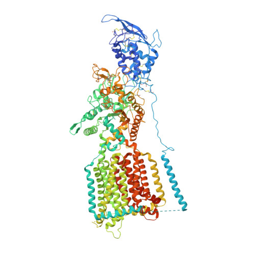
NPC1-NPC2 complex structure at pH 5.5


Experimental Data Snapshot

  • Method: ELECTRON MICROSCOPY
  • Resolution: 4.00 Å
  • Aggregation State: PARTICLE 
  • Reconstruction Method: SINGLE PARTICLE 

wwPDB Validation   3D Report Full Report


This is version 2.1 of the entry. See complete history


Literature

Structural Basis of Low-pH-Dependent Lysosomal Cholesterol Egress by NPC1 and NPC2.

Qian, H.Wu, X.Du, X.Yao, X.Zhao, X.Lee, J.Yang, H.Yan, N.

(2020) Cell 182: 98-111.e18

  • DOI: https://doi.org/10.1016/j.cell.2020.05.020
  • Primary Citation of Related Structures:  
    6W5R, 6W5S, 6W5T, 6W5U, 6W5V

  • PubMed Abstract: 

    Lysosomal cholesterol egress requires two proteins, NPC1 and NPC2, whose defects are responsible for Niemann-Pick disease type C (NPC). Here, we present systematic structural characterizations that reveal the molecular basis for low-pH-dependent cholesterol delivery from NPC2 to the transmembrane (TM) domain of NPC1. At pH 8.0, similar structures of NPC1 were obtained in nanodiscs and in detergent at resolutions of 3.6 Å and 3.0 Å, respectively. A tunnel connecting the N-terminal domain (NTD) and the transmembrane sterol-sensing domain (SSD) was unveiled. At pH 5.5, the NTD exhibits two conformations, suggesting the motion for cholesterol delivery to the tunnel. A putative cholesterol molecule is found at the membrane boundary of the tunnel, and TM2 moves toward formation of a surface pocket on the SSD. Finally, the structure of the NPC1-NPC2 complex at 4.0 Å resolution was obtained at pH 5.5, elucidating the molecular basis for cholesterol handoff from NPC2 to NPC1(NTD).


  • Organizational Affiliation

    Department of Molecular Biology, Princeton University, Princeton, NJ 08544, USA.


Macromolecules
Find similar proteins by:  (by identity cutoff)  |  3D Structure
Entity ID: 1
MoleculeChains Sequence LengthOrganismDetailsImage
NPC intracellular cholesterol transporter 11,311Homo sapiensMutation(s): 0 
Gene Names: NPC1
Membrane Entity: Yes 
UniProt & NIH Common Fund Data Resources
Find proteins for O15118 (Homo sapiens)
Explore O15118 
Go to UniProtKB:  O15118
PHAROS:  O15118
GTEx:  ENSG00000141458 
Entity Groups  
Sequence Clusters30% Identity50% Identity70% Identity90% Identity95% Identity100% Identity
UniProt GroupO15118
Glycosylation
Glycosylation Sites: 13Go to GlyGen: O15118-1
Sequence Annotations
Expand
  • Reference Sequence
Find similar proteins by:  (by identity cutoff)  |  3D Structure
Entity ID: 2
MoleculeChains Sequence LengthOrganismDetailsImage
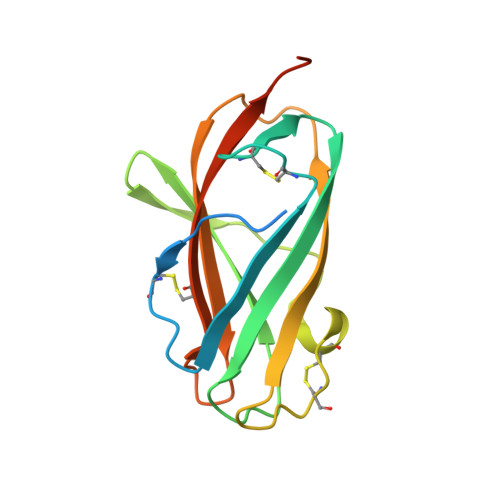
NPC intracellular cholesterol transporter 2B [auth D]161Homo sapiensMutation(s): 0 
Gene Names: NPC2HE1
Membrane Entity: Yes 
UniProt & NIH Common Fund Data Resources
Find proteins for P61916 (Homo sapiens)
Explore P61916 
Go to UniProtKB:  P61916
PHAROS:  P61916
GTEx:  ENSG00000119655 
Entity Groups  
Sequence Clusters30% Identity50% Identity70% Identity90% Identity95% Identity100% Identity
UniProt GroupP61916
Sequence Annotations
Expand
  • Reference Sequence
Oligosaccharides

Help

Entity ID: 3
MoleculeChains Length2D Diagram Glycosylation3D Interactions
2-acetamido-2-deoxy-beta-D-glucopyranose-(1-4)-2-acetamido-2-deoxy-beta-D-glucopyranoseC [auth B],
E,
F,
G
2N-Glycosylation
Glycosylation Resources
GlyTouCan:  G42666HT
GlyCosmos:  G42666HT
GlyGen:  G42666HT
Entity ID: 4
MoleculeChains Length2D Diagram Glycosylation3D Interactions
beta-D-mannopyranose-(1-3)-2-acetamido-2-deoxy-beta-D-glucopyranose-(1-4)-2-acetamido-2-deoxy-beta-D-glucopyranoseD [auth C]3N-Glycosylation
Glycosylation Resources
GlyTouCan:  G44266EX
GlyCosmos:  G44266EX
GlyGen:  G44266EX
Small Molecules
Ligands 2 Unique
IDChains Name / Formula / InChI Key2D Diagram3D Interactions
CLR (Subject of Investigation/LOI)
Query on CLR

Download Ideal Coordinates CCD File 
Q [auth A],
R [auth A],
T [auth D]
CHOLESTEROL
C27 H46 O
HVYWMOMLDIMFJA-DPAQBDIFSA-N
NAG
Query on NAG

Download Ideal Coordinates CCD File 
H [auth A]
I [auth A]
J [auth A]
K [auth A]
L [auth A]
H [auth A],
I [auth A],
J [auth A],
K [auth A],
L [auth A],
M [auth A],
N [auth A],
O [auth A],
P [auth A],
S [auth D]
2-acetamido-2-deoxy-beta-D-glucopyranose
C8 H15 N O6
OVRNDRQMDRJTHS-FMDGEEDCSA-N
Experimental Data & Validation

Experimental Data

  • Method: ELECTRON MICROSCOPY
  • Resolution: 4.00 Å
  • Aggregation State: PARTICLE 
  • Reconstruction Method: SINGLE PARTICLE 
EM Software:
TaskSoftware PackageVersion
RECONSTRUCTIONRELION

Structure Validation

View Full Validation Report


Entry History & Funding Information

Deposition Data


Funding OrganizationLocationGrant Number
National Science Foundation (NSF, United States)United StatesDMR-1420541

Revision History  (Full details and data files)

  • Version 1.0: 2020-06-17
    Type: Initial release
  • Version 1.1: 2020-07-01
    Changes: Database references
  • Version 1.2: 2020-07-22
    Changes: Database references
  • Version 2.0: 2020-07-29
    Type: Remediation
    Reason: Carbohydrate remediation
    Changes: Advisory, Atomic model, Data collection, Derived calculations, Structure summary
  • Version 2.1: 2024-10-23
    Changes: Data collection, Database references, Structure summary