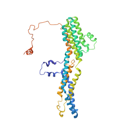
6VX9

bestrophin-2 Ca2+- unbound state 1 (EGTA only)


Experimental Data Snapshot

  • Method: ELECTRON MICROSCOPY
  • Resolution: 2.17 Å
  • Aggregation State: PARTICLE 
  • Reconstruction Method: SINGLE PARTICLE 

wwPDB Validation   3D Report Full Report


This is version 1.2 of the entry. See complete history


Literature

Structural and functional characterization of the bestrophin-2 anion channel.

Owji, A.P.Zhao, Q.Ji, C.Kittredge, A.Hopiavuori, A.Fu, Z.Ward, N.Clarke, O.B.Shen, Y.Zhang, Y.Hendrickson, W.A.Yang, T.

(2020) Nat Struct Mol Biol 27: 382-391

  • DOI: https://doi.org/10.1038/s41594-020-0402-z
  • Primary Citation of Related Structures:  
    6VX5, 6VX6, 6VX7, 6VX8, 6VX9

  • PubMed Abstract: 

    The bestrophin family of calcium (Ca 2+ )-activated chloride (Cl - ) channels, which mediate the influx and efflux of monovalent anions in response to the levels of intracellular Ca 2+ , comprises four members in mammals (bestrophin 1-4). Here we report cryo-EM structures of bovine bestrophin-2 (bBest2) bound and unbound by Ca 2+ at 2.4- and 2.2-Å resolution, respectively. The bBest2 structure highlights four previously underappreciated pore-lining residues specifically conserved in Best2 but not in Best1, illustrating the differences between these paralogs. Structure-inspired electrophysiological analysis reveals that, although the channel is sensitive to Ca 2+ , it has substantial Ca 2+ -independent activity for Cl - , reflecting the opening at the cytoplasmic restriction of the ion conducting pathway even when Ca 2+ is absent. Moreover, the ion selectivity of bBest2 is controlled by multiple residues, including those involved in gating.


  • Organizational Affiliation

    Department of Pharmacology, Columbia University, New York, NY, USA.


Macromolecules
Find similar proteins by:  (by identity cutoff)  |  3D Structure
Entity ID: 1
MoleculeChains Sequence LengthOrganismDetailsImage
BestrophinA,
B [auth E],
C [auth B],
D [auth C],
E [auth D]
410Bos taurusMutation(s): 0 
Gene Names: BEST2
Membrane Entity: Yes 
UniProt
Find proteins for E1BF86 (Bos taurus)
Explore E1BF86 
Go to UniProtKB:  E1BF86
Entity Groups  
Sequence Clusters30% Identity50% Identity70% Identity90% Identity95% Identity100% Identity
UniProt GroupE1BF86
Sequence Annotations
Expand
  • Reference Sequence
Small Molecules
Experimental Data & Validation

Experimental Data

  • Method: ELECTRON MICROSCOPY
  • Resolution: 2.17 Å
  • Aggregation State: PARTICLE 
  • Reconstruction Method: SINGLE PARTICLE 
EM Software:
TaskSoftware PackageVersion
RECONSTRUCTIONcryoSPARC2.13.2

Structure Validation

View Full Validation Report


Entry History & Funding Information

Deposition Data


Funding OrganizationLocationGrant Number
National Institutes of Health/National Eye Institute (NIH/NEI)United StatesF31EY030763
National Institutes of Health/National Eye Institute (NIH/NEI)United StatesEY025290
National Institutes of Health/National Institute of General Medical Sciences (NIH/NIGMS)United StatesR01GM127652
National Institutes of Health/National Institute of General Medical Sciences (NIH/NIGMS)United StatesR01GM107462

Revision History  (Full details and data files)

  • Version 1.0: 2020-04-08
    Type: Initial release
  • Version 1.1: 2020-04-22
    Changes: Database references
  • Version 1.2: 2024-03-06
    Changes: Data collection, Database references