
 Download Mendeley
A Positive Charge in the Outer Coordination Sphere of an Artificial Enzyme Increases CO2 Hydrogenation
Laureanti, J.A., Ginovska, B., Buchko, G.W., Schenter, G.K., Zadvornyy, O.A., Heberta, M., Peters, J.W., Shaw, W.J.(2020) Organometallics
Experimental Data Snapshot
Starting Model: experimental
View more details
(2020) Organometallics
Entity ID: 1 | |||||
|---|---|---|---|---|---|
| Molecule | Chains | Sequence Length | Organism | Details | Image |
| Transcriptional regulator, PadR-like family | 119 | Lactococcus cremoris subsp. cremoris MG1363 | Mutation(s): 4 Gene Names: llmg_0323 | 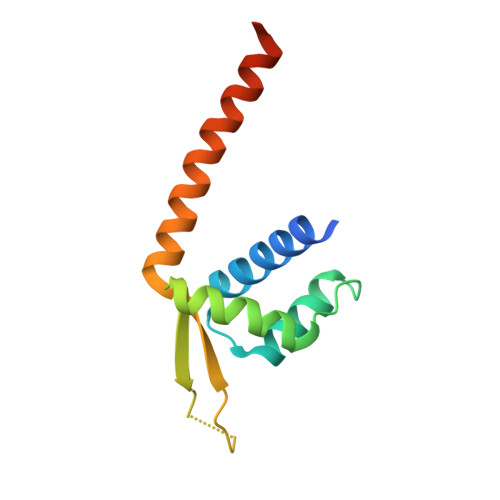 | |
UniProt | |||||
Entity Groups | |||||
| Sequence Clusters | 30% Identity50% Identity70% Identity90% Identity95% Identity100% Identity | ||||
| UniProt Group | A2RI36 | ||||
Sequence AnnotationsExpand | |||||
Reference Sequence | |||||
| Ligands 1 Unique | |||||
|---|---|---|---|---|---|
| ID | Chains | Name / Formula / InChI Key | 2D Diagram | 3D Interactions | |
| JY1 (Subject of Investigation/LOI) Download:Ideal Coordinates CCD File | C [auth A] | bis{N~2~,N~2~-bis[(diethylphosphanuidyl-kappaP)methyl]-N-[2-(2,5-dioxopyrrolidin-1-yl)ethyl]glycinamide}rhodium C36 H70 N6 O6 P4 Rh KIKWETXGDLBDBZ-UHFFFAOYSA-R | 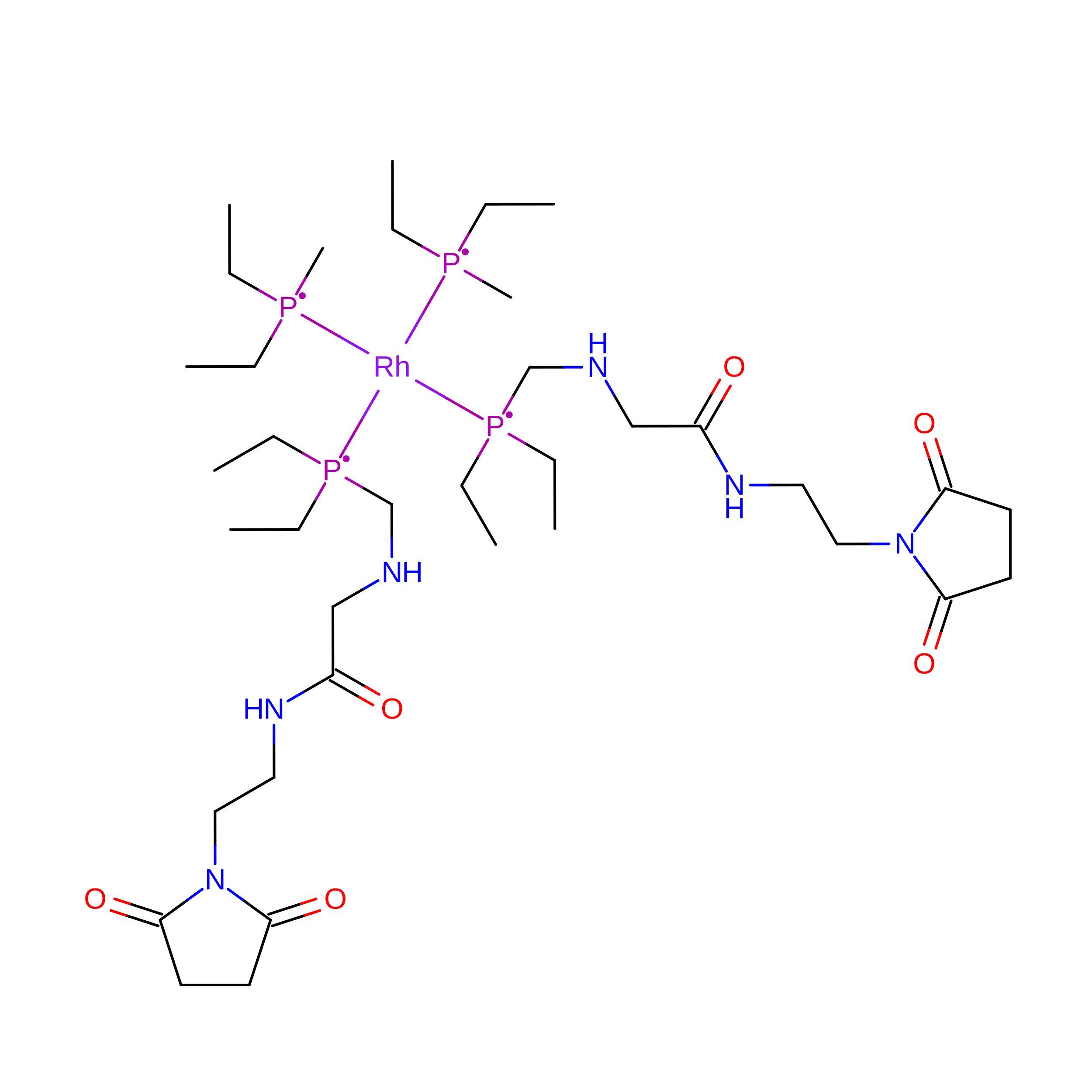 | ||
| Length ( Å ) | Angle ( ˚ ) |
|---|---|
| a = 101.855 | α = 90 |
| b = 35.619 | β = 95.92 |
| c = 71.651 | γ = 90 |
| Software Name | Purpose |
|---|---|
| PHENIX | refinement |
| XDS | data reduction |
| Aimless | data scaling |
| PHENIX | phasing |
| Funding Organization | Location | Grant Number |
|---|---|---|
| Department of Energy (DOE, United States) | United States | DE-FG02-04ER151563 |
| Department of Energy (DOE, United States) | United States | DE-AC02-76SF00515 |
| National Institutes of Health/National Institute of General Medical Sciences (NIH/NIGMS) | United States | P41GM103393 |
