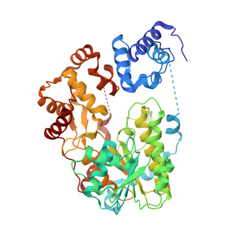
6VTJ

Structure of a thiolation-reductase di-domain from an archaeal non-ribosomal peptide synthetase


Experimental Data Snapshot

  • Method: X-RAY DIFFRACTION
  • Resolution: 1.95 Å
  • R-Value Free: 0.239 
  • R-Value Work: 0.196 
  • R-Value Observed: 0.198 

Starting Model: experimental
View more details

wwPDB Validation   3D Report Full Report

Currently 6VTJ does not have a validation slider image.


This is version 2.3 of the entry. See complete history


Literature

Structural characterization of a PCP-R didomain from an archaeal nonribosomal peptide synthetase reveals novel interdomain interactions.

Deshpande, S.Altermann, E.Sarojini, V.Lott, J.S.Lee, T.V.

(2021) J Biol Chem 296: 100432-100432

  • DOI: https://doi.org/10.1016/j.jbc.2021.100432
  • Primary Citation of Related Structures:  
    6VTJ, 6VTZ

  • PubMed Abstract: 

    Nonribosomal peptide synthetases (NRPSs) are multimodular enzymes that produce a wide range of bioactive peptides, such as siderophores, toxins, and antibacterial and insecticidal agents. NRPSs are dynamic proteins characterized by extensive interdomain communications as a consequence of their assembly-line mode of synthesis. Hence, crystal structures of multidomain fragments of NRPSs have aided in elucidating crucial interdomain interactions that occur during different steps of the NRPS catalytic cycle. One crucial yet unexplored interaction is that between the reductase (R) domain and the peptide carrier protein (PCP) domain. R domains are members of the short-chain dehydrogenase/reductase family and function as termination domains that catalyze the reductive release of the final peptide product from the terminal PCP domain of the NRPS. Here, we report the crystal structure of an archaeal NRPS PCP-R didomain construct. This is the first NRPS R domain structure to be determined together with the upstream PCP domain and is also the first structure of an archaeal NRPS to be reported. The structure reveals that a novel helix-turn-helix motif, found in NRPS R domains but not in other short-chain dehydrogenase/reductase family members, plays a major role in the interface between the PCP and R domains. The information derived from the described PCP-R interface will aid in gaining further mechanistic insights into the peptide termination reaction catalyzed by the R domain and may have implications in engineering NRPSs to synthesize novel peptide products.


  • Organizational Affiliation

    School of Biological Sciences, University of Auckland, Auckland, New Zealand.


Macromolecules
Find similar proteins by:  (by identity cutoff)  |  3D Structure
Entity ID: 1
MoleculeChains Sequence LengthOrganismDetailsImage
Non-ribosomal peptide synthetase491Methanobrevibacter ruminantium M1Mutation(s): 0 
Gene Names: mru_0351
UniProt
Find proteins for D3E027 (Methanobrevibacter ruminantium (strain ATCC 35063 / DSM 1093 / JCM 13430 / OCM 146 / M1))
Explore D3E027 
Go to UniProtKB:  D3E027
Entity Groups  
Sequence Clusters30% Identity50% Identity70% Identity90% Identity95% Identity100% Identity
UniProt GroupD3E027
Sequence Annotations
Expand
  • Reference Sequence
Small Molecules
Ligands 1 Unique
IDChains Name / Formula / InChI Key2D Diagram3D Interactions
PNS (Subject of Investigation/LOI)
Query on PNS

Download Ideal Coordinates CCD File 
B [auth A]4'-PHOSPHOPANTETHEINE
C11 H23 N2 O7 P S
JDMUPRLRUUMCTL-VIFPVBQESA-N
Experimental Data & Validation

Experimental Data

  • Method: X-RAY DIFFRACTION
  • Resolution: 1.95 Å
  • R-Value Free: 0.239 
  • R-Value Work: 0.196 
  • R-Value Observed: 0.198 
  • Space Group: C 1 2 1
Unit Cell:
Length ( Å )Angle ( ˚ )
a = 98.949α = 90
b = 71.833β = 90.26
c = 82.725γ = 90
Software Package:
Software NamePurpose
PHENIXrefinement
XDSdata reduction
Aimlessdata scaling
PDB_EXTRACTdata extraction
PHASERphasing

Structure Validation

View Full Validation Report

Currently 6VTJ does not have a validation slider image.


Entry History & Funding Information

Deposition Data


Funding OrganizationLocationGrant Number
Ministry of Business, Innovation and Employment (New Zealand)New ZealandUOAX1506

Revision History  (Full details and data files)

  • Version 1.0: 2020-02-26
    Type: Initial release
  • Version 2.0: 2021-03-10
    Type: Coordinate replacement
    Reason: Ligand geometry
    Changes: Atomic model, Data collection, Derived calculations, Experimental preparation, Refinement description
  • Version 2.1: 2021-03-17
    Changes: Database references
  • Version 2.2: 2021-07-21
    Changes: Database references
  • Version 2.3: 2023-10-11
    Changes: Data collection, Database references, Refinement description