6VSI

Crystal structure of FKBP12 of Candida auris


Experimental Data Snapshot

  • Method: X-RAY DIFFRACTION
  • Resolution: 1.87 Å
  • R-Value Free: 0.206 
  • R-Value Work: 0.202 
  • R-Value Observed: 0.202 

wwPDB Validation   3D Report Full Report

Currently 6VSI does not have a validation slider image.


This is version 1.2 of the entry. See complete history


Literature

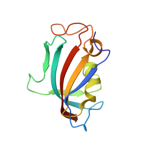
Crystal structure and transient dimerization for the FKBP12 protein from the pathogenic fungus Candida auris.

Bashir, Q.Li, Z.Li, H.LeMaster, D.M.Hernandez, G.

(2020) Biochem Biophys Res Commun 525: 1103-1108

  • DOI: https://doi.org/10.1016/j.bbrc.2020.03.059
  • Primary Citation of Related Structures:  
    6VSI

  • PubMed Abstract: 

    International concern over the recent emergence of Candida auris infections reflects not only its comparative ease of transmission and substantial mortality but the increasing level of resistance observed to all three major classes of antifungal drugs. Diminution in virulence has been reported for a wide range of fungal pathogens when the FK506-binding protein FKBP12 binds to that immunosuppressant drug and the binary complex then inhibits the fungal calcineurin signaling pathway. Structure-based drug design efforts have described modifications of FK506 which modestly reduce virulence for a number of fungal pathogens while also lessening the side effect of suppressing the tissue immunity response in the patient. To aid in such studies, we report the crystal structure of Candida auris FKBP12. As physiological relevance has been proposed for transient homodimerization interactions of distantly related fungal FKBP12 proteins, we report the solution NMR characterization of the homodimerization interactions of the FKBP12 proteins from both Candida auris and Candida glabrata.


  • Organizational Affiliation

    Wadsworth Center, New York State Department of Health, Empire State Plaza, Albany, NY, 12201, USA.


Macromolecules
Find similar proteins by:  (by identity cutoff)  |  3D Structure
Entity ID: 1
MoleculeChains Sequence LengthOrganismDetailsImage
Peptidylprolyl isomerase111[Candida] aurisMutation(s): 0 
Gene Names: CJJ09_002997
EC: 5.2.1.8
UniProt
Find proteins for A0A2H1A4Z6 (Candida auris)
Explore A0A2H1A4Z6 
Go to UniProtKB:  A0A2H1A4Z6
Entity Groups  
Sequence Clusters30% Identity50% Identity70% Identity90% Identity95% Identity100% Identity
UniProt GroupA0A2H1A4Z6
Sequence Annotations
Expand
  • Reference Sequence
Small Molecules
Ligands 1 Unique
IDChains Name / Formula / InChI Key2D Diagram3D Interactions
SO4
Query on SO4

Download Ideal Coordinates CCD File 
B [auth A]SULFATE ION
O4 S
QAOWNCQODCNURD-UHFFFAOYSA-L
Experimental Data & Validation

Experimental Data

  • Method: X-RAY DIFFRACTION
  • Resolution: 1.87 Å
  • R-Value Free: 0.206 
  • R-Value Work: 0.202 
  • R-Value Observed: 0.202 
  • Space Group: P 43 2 2
Unit Cell:
Length ( Å )Angle ( ˚ )
a = 74.8α = 90
b = 74.8β = 90
c = 72.29γ = 90
Software Package:
Software NamePurpose
XSCALEdata scaling
PHASERphasing
PHENIXrefinement
PDB_EXTRACTdata extraction
XDSdata reduction

Structure Validation

View Full Validation Report

Currently 6VSI does not have a validation slider image.


Entry History & Funding Information

Deposition Data


Funding OrganizationLocationGrant Number
National Institutes of Health/National Institute of General Medical Sciences (NIH/NIGMS)United StatesGM119152

Revision History  (Full details and data files)

  • Version 1.0: 2020-04-01
    Type: Initial release
  • Version 1.1: 2020-04-29
    Changes: Database references
  • Version 1.2: 2023-10-11
    Changes: Advisory, Data collection, Database references, Refinement description