6VLF | pdb_00006vlf

Crystal structure of mouse alpha 1,6-fucosyltransferase, FUT8 in its Apo-form


Experimental Data Snapshot

  • Method: X-RAY DIFFRACTION
  • Resolution: 1.80 Å
  • R-Value Free: 
    0.208 (Depositor), 0.209 (DCC) 
  • R-Value Work: 
    0.184 (Depositor), 0.185 (DCC) 
  • R-Value Observed: 
    0.185 (Depositor) 

Starting Model: experimental
View more details

wwPDB Validation 3D Report Full Report

Validation slider image for 6VLF

This is version 1.3 of the entry. See complete history

Literature

Structural basis of substrate recognition and catalysis by fucosyltransferase 8.

Jarva, M.A.Dramicanin, M.Lingford, J.P.Mao, R.John, A.Jarman, K.E.Grinter, R.Goddard-Borger, E.D.

(2020) J Biological Chem 295: 6677-6688

  • DOI: https://doi.org/10.1074/jbc.RA120.013291
  • Primary Citation Related Structures: 
    6VLD, 6VLE, 6VLF, 6VLG

  • PubMed Abstract: 

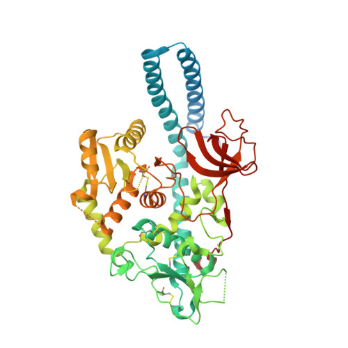
    Fucosylation of the innermost GlcNAc of N -glycans by fucosyltransferase 8 (FUT8) is an important step in the maturation of complex and hybrid N -glycans. This simple modification can dramatically affect the activities and half-lives of glycoproteins, effects that are relevant to understanding the invasiveness of some cancers, development of mAb therapeutics, and the etiology of a congenital glycosylation disorder. The acceptor substrate preferences of FUT8 are well-characterized and provide a framework for understanding N -glycan maturation in the Golgi; however, the structural basis of these substrate preferences and the mechanism through which catalysis is achieved remain unknown. Here we describe several structures of mouse and human FUT8 in the apo state and in complex with GDP, a mimic of the donor substrate, and with a glycopeptide acceptor substrate at 1.80-2.50 Å resolution. These structures provide insights into a unique conformational change associated with donor substrate binding, common strategies employed by fucosyltransferases to coordinate GDP, features that define acceptor substrate preferences, and a likely mechanism for enzyme catalysis. Together with molecular dynamics simulations, the structures also revealed how FUT8 dimerization plays an important role in defining the acceptor substrate-binding site. Collectively, this information significantly builds on our understanding of the core fucosylation process.


  • Organizational Affiliation
    • The Walter and Eliza Hall Institute of Medical Research, Parkville, Victoria 3052, Australia.

Macromolecule Content 

  • Total Structure Weight: 126.15 kDa 
  • Atom Count: 8,066 
  • Modeled Residue Count: 898 
  • Deposited Residue Count: 1,086 
  • Unique protein chains: 1

Macromolecules

Find similar proteins by:|  3D Structure
Entity ID: 1
MoleculeChains  Sequence LengthOrganismDetailsImage
Alpha-(1,6)-fucosyltransferase
A, B
543Mus musculusMutation(s): 0 
Gene Names: Fut8
EC: 2.4.1.68
UniProt
Find proteins for Q9WTS2 (Mus musculus)
Explore Q9WTS2 
Go to UniProtKB:  Q9WTS2
Entity Groups
Sequence Clusters30% Identity50% Identity70% Identity90% Identity95% Identity100% Identity
UniProt GroupQ9WTS2
Sequence Annotations
Expand
Reference Sequence

Small Molecules

Ligands 2 Unique
IDChains Name / Formula / InChI Key2D Diagram3D Interactions
SO4

Query on SO4


Download:Ideal Coordinates CCD File
C [auth A],
D [auth A],
K [auth B],
L [auth B]
SULFATE ION
O4 S
QAOWNCQODCNURD-UHFFFAOYSA-L
EDO

Query on EDO


Download:Ideal Coordinates CCD File
E [auth A]
F [auth A]
G [auth A]
H [auth A]
I [auth A]
E [auth A],
F [auth A],
G [auth A],
H [auth A],
I [auth A],
J [auth A],
M [auth B],
N [auth B],
O [auth B]
1,2-ETHANEDIOL
C2 H6 O2
LYCAIKOWRPUZTN-UHFFFAOYSA-N

Experimental Data & Validation

Experimental Data

  • Method: X-RAY DIFFRACTION
  • Resolution: 1.80 Å
  • R-Value Free:  0.208 (Depositor), 0.209 (DCC) 
  • R-Value Work:  0.184 (Depositor), 0.185 (DCC) 
  • R-Value Observed: 0.185 (Depositor) 
Space Group: C 1 2 1
Unit Cell:
Length ( Å )Angle ( ˚ )
a = 184.874α = 90
b = 71.353β = 126.08
c = 126.951γ = 90
Software Package:
Software NamePurpose
PHENIXrefinement
XDSdata reduction
Aimlessdata scaling
PHASERphasing
PDB_EXTRACTdata extraction

Structure Validation

View Full Validation Report


Entry History 

& Funding Information

Deposition Data


Funding OrganizationLocationGrant Number
Other governmentAustraliaGNT1139546

Revision History  (Full details and data files)

  • Version 1.0: 2020-02-26
    Type: Initial release
  • Version 1.1: 2020-09-16
    Changes: Database references
  • Version 1.2: 2023-10-11
    Changes: Data collection, Database references, Refinement description
  • Version 1.3: 2024-10-16
    Changes: Structure summary