6VJV | pdb_00006vjv

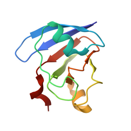
Crystal structure of the Prochlorococcus phage (myovirus P-SSM2) ferredoxin at 1.6 Angstroms


Experimental Data Snapshot

  • Method: X-RAY DIFFRACTION
  • Resolution: 1.59 Å
  • R-Value Free: 
    0.213 (Depositor), 0.219 (DCC) 
  • R-Value Work: 
    0.178 (Depositor), 0.181 (DCC) 
  • R-Value Observed: 
    0.180 (Depositor) 

Starting Model: experimental
View more details

wwPDB Validation 3D Report Full Report

Validation slider image for 6VJV

This is version 1.4 of the entry. See complete history. 

Literature

Prochlorococcusphage ferredoxin: structural characterization and electron transfer to cyanobacterial sulfite reductases.

Campbell, I.J., Olmos Jr., J.L., Xu, W., Kahanda, D., Atkinson, J.T., Sparks, O.N., Miller, M.D., Phillips Jr., G.N., Bennett, G.N., Silberg, J.J.

(2020) J Biol Chem 295: 10610-10623

  • DOI: https://doi.org/10.1074/jbc.RA120.013501
  • Primary Citation Related Structures: 
    6VJV

  • PubMed Abstract: 

    Marine cyanobacteria are infected by phages whose genomes encode ferredoxin (Fd) electron carriers. These Fds are thought to redirect the energy harvested from light to phage-encoded oxidoreductases that enhance viral fitness, but it is unclear how the biophysical properties and partner specificities of phage Fds relate to those of photosynthetic organisms. Here, results of a bioinformatics analysis using a sequence similarity network revealed that phage Fds are most closely related to cyanobacterial Fds that transfer electrons from photosystems to oxidoreductases involved in nutrient assimilation. Structural analysis of myovirus P-SSM2 Fd (pssm2-Fd), which infects the cyanobacterium Prochlorococcus marinus , revealed high levels of similarity to cyanobacterial Fds (root mean square deviations of ≤0.5 Å). Additionally, pssm2-Fd exhibited a low midpoint reduction potential (-336 mV versus a standard hydrogen electrode), similar to other photosynthetic Fds, although it had lower thermostability ( T m = 28 °C) than did many other Fds. When expressed in an Escherichia coli strain deficient in sulfite assimilation, pssm2-Fd complemented bacterial growth when coexpressed with a P. marinus sulfite reductase, revealing that pssm2-Fd can transfer electrons to a host protein involved in nutrient assimilation. The high levels of structural similarity with cyanobacterial Fds and reactivity with a host sulfite reductase suggest that phage Fds evolved to transfer electrons to cyanobacterially encoded oxidoreductases.


  • Organizational Affiliation: 
    • Biochemistry and Cell Biology Graduate Program, Rice University, Houston, Texas, USA.

Macromolecule Content 

  • Total Structure Weight: 22.27 kDa 
  • Atom Count: 1,757 
  • Modeled Residue Count: 192 
  • Deposited Residue Count: 192 
  • Unique protein chains: 1

Macromolecules

Find similar proteins by:|  3D Structure
Entity ID: 1
MoleculeChains  Sequence LengthOrganismDetailsImage
Ferredoxin
A, B
96Prochlorococcus phage P-SSM2Mutation(s): 0 
Gene Names: PCMG_00283, PSSM2_281
UniProt
Find proteins for Q58M74 (Prochlorococcus phage P-SSM2)
Explore Q58M74 
Go to UniProtKB:  Q58M74
Entity Groups
Sequence Clusters30% Identity50% Identity70% Identity90% Identity95% Identity100% Identity
UniProt GroupQ58M74
Sequence Annotations
Expand
Reference Sequence

Small Molecules

Ligands 3 Unique
IDChains Name / Formula / InChI Key2D Diagram3D Interactions
FES
(Subject of Investigation/LOI)

Query on FES


Download:Ideal Coordinates CCD File
C [auth A],
M [auth B]
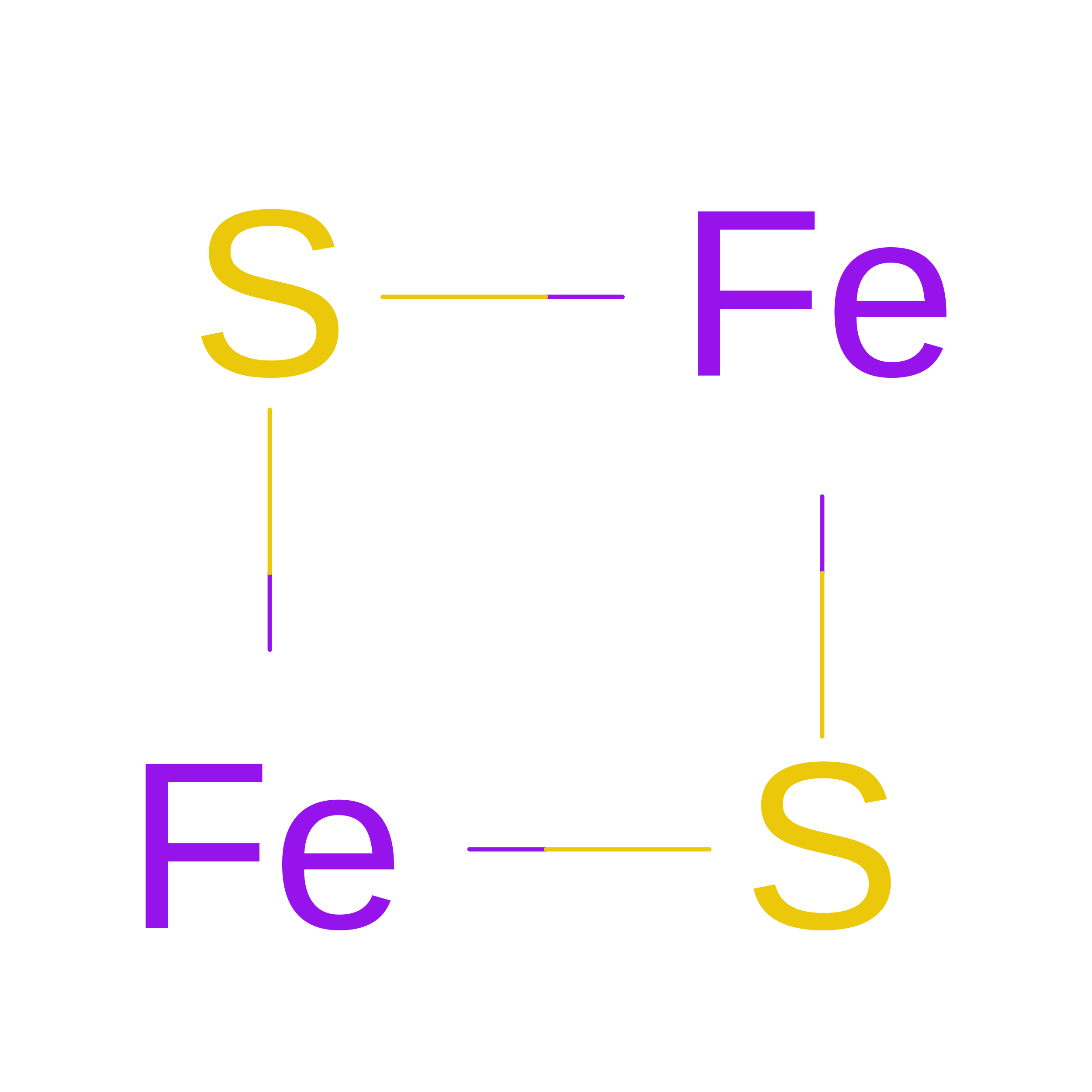
FE2/S2 (INORGANIC) CLUSTER
Fe2 S2
NIXDOXVAJZFRNF-UHFFFAOYSA-N
ZN

Query on ZN


Download:Ideal Coordinates CCD File
F [auth A]
G [auth A]
H [auth A]
I [auth A]
J [auth A]
F [auth A],
G [auth A],
H [auth A],
I [auth A],
J [auth A],
K [auth A],
L [auth A],
P [auth B],
Q [auth B],
R [auth B],
S [auth B],
T [auth B],
U [auth B],
V [auth B]
ZINC ION
Zn
PTFCDOFLOPIGGS-UHFFFAOYSA-N
ACT

Query on ACT


Download:Ideal Coordinates CCD File
D [auth A],
E [auth A],
N [auth B],
O [auth B]
ACETATE ION
C2 H3 O2
QTBSBXVTEAMEQO-UHFFFAOYSA-M

Experimental Data & Validation

Experimental Data

  • Method: X-RAY DIFFRACTION
  • Resolution: 1.59 Å
  • R-Value Free:  0.213 (Depositor), 0.219 (DCC) 
  • R-Value Work:  0.178 (Depositor), 0.181 (DCC) 
  • R-Value Observed: 0.180 (Depositor) 
Space Group: P 1 21 1
Unit Cell:
Length ( Å )Angle ( ˚ )
a = 52.619α = 90
b = 31.446β = 92.48
c = 61.288γ = 90
Software Package:
Software NamePurpose
PHENIXrefinement
XDSdata reduction
Aimlessdata scaling
PHASERphasing

Structure Validation

View Full Validation Report


Entry History 

& Funding Information

Deposition Data


Funding OrganizationLocationGrant Number
Department of Energy (DOE, United States)United StatesDE-SC0014462
National Science Foundation (NSF, United States)United States1231306
National Aeronautic Space Administration (NASA, United States)United States80NSSC18M0093

Revision History  (Full details and data files)

  • Version 1.0: 2020-02-19
    Type: Initial release
  • Version 1.1: 2020-06-03
    Changes: Database references
  • Version 1.2: 2020-08-12
    Changes: Database references, Derived calculations
  • Version 1.3: 2023-10-11
    Changes: Data collection, Database references, Refinement description
  • Version 1.4: 2026-08-12
    Type: Remediation
    Reason: Metalloprotein remediation
    Changes: Advisory, Derived calculations, Structure summary