6VJI | pdb_00006vji

Structure of mammalian NEIL2 from Monodelphis domestica


Experimental Data Snapshot

  • Method: X-RAY DIFFRACTION
  • Resolution: 2.54 Å
  • R-Value Free: 
    0.275 (Depositor), 0.277 (DCC) 
  • R-Value Work: 
    0.250 (Depositor), 0.255 (DCC) 
  • R-Value Observed: 
    0.253 (Depositor) 

wwPDB Validation 3D Report Full Report

Validation slider image for 6VJI

This is version 1.3 of the entry. See complete history

Literature

Unique Structural Features of Mammalian NEIL2 DNA Glycosylase Prime Its Activity for Diverse DNA Substrates and Environments.

Eckenroth, B.E.Cao, V.B.Averill, A.M.Dragon, J.A.Doublie, S.

(2021) Structure 29: 29-42.e4

  • DOI: https://doi.org/10.1016/j.str.2020.08.001
  • Primary Citation Related Structures: 
    6VJI

  • PubMed Abstract: 

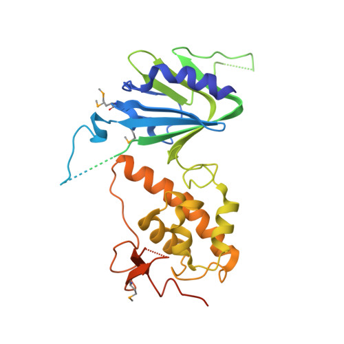
    Oxidative damage on DNA arising from both endogenous and exogenous sources can result in base modifications that promote errors in replication as well as generating sites of base loss (abasic sites) that present unique challenges to maintaining genomic integrity. These lesions are excised by DNA glycosylases in the first step of the base excision repair pathway. Here we present the first crystal structure of a NEIL2 glycosylase, an enzyme active on cytosine oxidation products and abasic sites. The structure reveals an unusual "open" conformation not seen in NEIL1 or NEIL3 orthologs. NEIL2 is predicted to adopt a "closed" conformation when bound to its substrate. Combined crystallographic and solution-scattering studies show the enzyme to be conformationally dynamic in a manner distinct among the NEIL glycosylases and provide insight into the unique substrate preference of this enzyme. In addition, we characterized three cancer variants of human NEIL2, namely S140N, G230W, and G303R.


  • Organizational Affiliation
    • Department of Microbiology and Molecular Genetics, University of Vermont, Stafford Hall, 95 Carrigan Drive, Burlington, VT 05405, USA. Electronic address: beckenro@uvm.edu.

Macromolecule Content 

  • Total Structure Weight: 79 kDa 
  • Atom Count: 3,815 
  • Modeled Residue Count: 508 
  • Deposited Residue Count: 688 
  • Unique protein chains: 1

Macromolecules

Find similar proteins by:|  3D Structure
Entity ID: 1
MoleculeChains  Sequence LengthOrganismDetailsImage
Nei like DNA glycosylase 2
A, B
344Monodelphis domesticaMutation(s): 0 
Gene Names: NEIL2
EC: 4.2.99.18
UniProt
Find proteins for F7AMK3 (Monodelphis domestica)
Explore F7AMK3 
Go to UniProtKB:  F7AMK3
Entity Groups
Sequence Clusters30% Identity50% Identity70% Identity90% Identity95% Identity100% Identity
UniProt GroupF7AMK3
Sequence Annotations
Expand
Reference Sequence

Small Molecules

Modified Residues  1 Unique
IDChains TypeFormula2D DiagramParent
MSE
Query on MSE
A, B
L-PEPTIDE LINKINGC5 H11 N O2 SeMET

Experimental Data & Validation

Experimental Data

  • Method: X-RAY DIFFRACTION
  • Resolution: 2.54 Å
  • R-Value Free:  0.275 (Depositor), 0.277 (DCC) 
  • R-Value Work:  0.250 (Depositor), 0.255 (DCC) 
  • R-Value Observed: 0.253 (Depositor) 
Space Group: P 32
Unit Cell:
Length ( Å )Angle ( ˚ )
a = 67.887α = 90
b = 67.887β = 90
c = 149.13γ = 120
Software Package:
Software NamePurpose
PHENIXrefinement
XDSdata reduction
Aimlessdata scaling
SHARPphasing
SOLOMONphasing
PDB_EXTRACTdata extraction

Structure Validation

View Full Validation Report


Entry History 

& Funding Information

Deposition Data


Funding OrganizationLocationGrant Number
National Institutes of Health/National Cancer Institute (NIH/NCI)United StatesCA098993
National Institutes of Health/National Cancer Institute (NIH/NCI)United StatesCA233185

Revision History  (Full details and data files)

  • Version 1.0: 2020-08-12
    Type: Initial release
  • Version 1.1: 2020-09-09
    Changes: Database references
  • Version 1.2: 2021-01-20
    Changes: Database references
  • Version 1.3: 2024-11-06
    Changes: Data collection, Database references, Refinement description, Structure summary