6VI8 | pdb_00006vi8

Observing a ring-cleaving dioxygenase in action through a crystalline lens - a superoxo bound structure


Experimental Data Snapshot

  • Method: X-RAY DIFFRACTION
  • Resolution: 1.95 Å
  • R-Value Free: 
    0.263 (Depositor), 0.239 (DCC) 
  • R-Value Work: 
    0.208 (Depositor) 
  • R-Value Observed: 
    0.214 (Depositor) 

Starting Model: experimental
View more details

wwPDB Validation 3D Report Full Report

Validation slider image for 6VI8

Ligand Structure Quality Assessment 


This is version 1.2 of the entry. See complete history

Literature

Observing 3-hydroxyanthranilate-3,4-dioxygenase in action through a crystalline lens.

Wang, Y.Liu, K.F.Yang, Y.Davis, I.Liu, A.

(2020) Proc Natl Acad Sci U S A 117: 19720-19730

  • DOI: https://doi.org/10.1073/pnas.2005327117
  • Primary Citation Related Structures: 
    6VI5, 6VI6, 6VI7, 6VI8, 6VI9, 6VIA, 6VIB, 6X11

  • PubMed Abstract: 

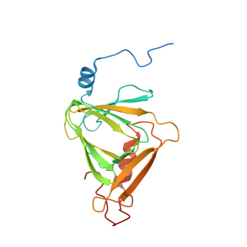
    The synthesis of quinolinic acid from tryptophan is a critical step in the de novo biosynthesis of nicotinamide adenine dinucleotide (NAD + ) in mammals. Herein, the nonheme iron-based 3-hydroxyanthranilate-3,4-dioxygenase responsible for quinolinic acid production was studied by performing time-resolved in crystallo reactions monitored by UV-vis microspectroscopy, electron paramagnetic resonance (EPR) spectroscopy, and X-ray crystallography. Seven catalytic intermediates were kinetically and structurally resolved in the crystalline state, and each accompanies protein conformational changes at the active site. Among them, a monooxygenated, seven-membered lactone intermediate as a monodentate ligand of the iron center at 1.59-Å resolution was captured, which presumably corresponds to a substrate-based radical species observed by EPR using a slurry of small-sized single crystals. Other structural snapshots determined at around 2.0-Å resolution include monodentate and subsequently bidentate coordinated substrate, superoxo, alkylperoxo, and two metal-bound enol tautomers of the unstable dioxygenase product. These results reveal a detailed stepwise O-atom transfer dioxygenase mechanism along with potential isomerization activity that fine-tunes product profiling and affects the production of quinolinic acid at a junction of the metabolic pathway.


  • Organizational Affiliation
    • Department of Chemistry, The University of Texas at San Antonio, San Antonio, TX 78249.

Macromolecule Content 

  • Total Structure Weight: 23.01 kDa 
  • Atom Count: 1,642 
  • Modeled Residue Count: 176 
  • Deposited Residue Count: 195 
  • Unique protein chains: 1

Macromolecules

Find similar proteins by:|  3D Structure
Entity ID: 1
MoleculeChains  Sequence LengthOrganismDetailsImage
3-hydroxyanthranilate 3,4-dioxygenase195Cupriavidus metallidurans CH34Mutation(s): 0 
Gene Names: nbaCRmet_5193
EC: 1.13.11.6
UniProt
Find proteins for Q1LCS4 (Cupriavidus metallidurans (strain ATCC 43123 / DSM 2839 / NBRC 102507 / CH34))
Explore Q1LCS4 
Go to UniProtKB:  Q1LCS4
Entity Groups
Sequence Clusters30% Identity50% Identity70% Identity90% Identity95% Identity100% Identity
UniProt GroupQ1LCS4
Sequence Annotations
Expand
Reference Sequence

Small Molecules

Ligands 5 Unique
IDChains Name / Formula / InChI Key2D Diagram3D Interactions
3HA
(Subject of Investigation/LOI)

Query on 3HA


Download:Ideal Coordinates CCD File
G [auth A]3-HYDROXYANTHRANILIC ACID
C7 H7 N O3
WJXSWCUQABXPFS-UHFFFAOYSA-N
TRS

Query on TRS


Download:Ideal Coordinates CCD File
B [auth A]2-AMINO-2-HYDROXYMETHYL-PROPANE-1,3-DIOL
C4 H12 N O3
LENZDBCJOHFCAS-UHFFFAOYSA-O
FE2

Query on FE2


Download:Ideal Coordinates CCD File
D [auth A]FE (II) ION
Fe
CWYNVVGOOAEACU-UHFFFAOYSA-N
FE
(Subject of Investigation/LOI)

Query on FE


Download:Ideal Coordinates CCD File
C [auth A]FE (III) ION
Fe
VTLYFUHAOXGGBS-UHFFFAOYSA-N
O
(Subject of Investigation/LOI)

Query on O


Download:Ideal Coordinates CCD File
E [auth A],
F [auth A]
OXYGEN ATOM
O
XLYOFNOQVPJJNP-UHFFFAOYSA-N

Experimental Data & Validation

Experimental Data

  • Method: X-RAY DIFFRACTION
  • Resolution: 1.95 Å
  • R-Value Free:  0.263 (Depositor), 0.239 (DCC) 
  • R-Value Work:  0.208 (Depositor) 
  • R-Value Observed: 0.214 (Depositor) 
Space Group: P 65 2 2
Unit Cell:
Length ( Å )Angle ( ˚ )
a = 58.04α = 90
b = 58.04β = 90
c = 231.29γ = 120
Software Package:
Software NamePurpose
PHENIXrefinement
DENZOdata reduction
SCALEPACKdata scaling
PDB_EXTRACTdata extraction
PHENIXphasing

Structure Validation

View Full Validation Report


Ligand Structure Quality Assessment 


Entry History 

& Funding Information

Deposition Data


Funding OrganizationLocationGrant Number
National Institutes of Health/National Institute of General Medical Sciences (NIH/NIGMS)United StatesR01GM108988
National Institutes of Health/National Institute of Mental Health (NIH/NIMH)United StatesR21MH107985
National Science Foundation (NSF, United States)United StatesCHE-1623856
National Institutes of Health/National Institute of General Medical Sciences (NIH/NIGMS)United StatesR01GM107529

Revision History  (Full details and data files)

  • Version 1.0: 2020-07-29
    Type: Initial release
  • Version 1.1: 2021-10-06
    Changes: Database references
  • Version 1.2: 2023-10-11
    Changes: Data collection, Refinement description