Experimental Phasing of MicroED Data Using Radiation Damage.
Martynowycz, M.W., Hattne, J., Gonen, T.(2020) Structure 28: 458
- PubMed: 32023481 Search on PubMedSearch on PubMed Central
- DOI: https://doi.org/10.1016/j.str.2020.01.008
- Primary Citation Related Structures:
6VHB, 6VHC - PubMed Abstract:
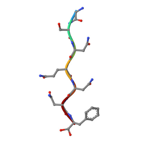
We previously demonstrated that microcrystal electron diffraction (MicroED) can be used to determine atomic-resolution structures from vanishingly small three-dimensional crystals. Here, we present an example of an experimentally phased structure using only MicroED data. The structure of a seven-residue peptide is solved starting from differences to the diffraction intensities induced by structural changes due to radiation damage. The same wedge of reciprocal space was recorded twice by continuous-rotation MicroED from a set of 11 individual crystals. The data from the first pass were merged to make a "low-dose dataset." The data from the second pass were similarly merged to form a "damaged dataset." Differences between these two datasets were used to identify a single heavy-atom site from a Patterson difference map, and initial phases were generated. Finally, the structure was completed by iterative cycles of modeling and refinement.
- Howard Hughes Medical Institute, Departments of Biological Chemistry and Physiology, David Geffen School of Medicine, University of California Los Angeles, Los Angeles, CA 90095, USA.
Organizational Affiliation: