6VEO | pdb_00006veo

ATAD2B bromodomain in complex with 4-({[(3R,4R)-4-{[3-methyl-5-(5-methylpyridin-3-yl)-2-oxo-1,2-dihydro-1,7-naphthyridin-8-yl]amino}piperidin-3-yl]oxy}methyl)-1lambda~6~-thiane-1,1-dione (compound 38)

  • Classification: NUCLEAR PROTEIN
  • Organism(s): Homo sapiens
  • Expression System: Escherichia coli
  • Mutation(s): No 

  • Deposited: 2020-01-02 Released: 2020-10-28 
  • Deposition Author(s): Glass, K.C.
  • Funding Organization(s): National Institutes of Health/National Institute of General Medical Sciences (NIH/NIGMS)

Experimental Data Snapshot

  • Method: X-RAY DIFFRACTION
  • Resolution: 2.40 Å
  • R-Value Free: 
    0.251 (Depositor), 0.252 (DCC) 
  • R-Value Work: 
    0.194 (Depositor), 0.194 (DCC) 
  • R-Value Observed: 
    0.197 (Depositor) 

Starting Model: experimental
View more details

wwPDB Validation 3D Report Full Report

Validation slider image for 6VEO

Ligand Structure Quality Assessment 


This is version 1.2 of the entry. See complete history

Literature

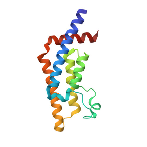
Structural Insights into the Recognition of Mono- and Diacetylated Histones by the ATAD2B Bromodomain.

Lloyd, J.T.McLaughlin, K.Lubula, M.Y.Gay, J.C.Dest, A.Gao, C.Phillips, M.Tonelli, M.Cornilescu, G.Marunde, M.R.Evans, C.M.Boyson, S.P.Carlson, S.Keogh, M.C.Markley, J.L.Frietze, S.Glass, K.C.

(2020) J Med Chem 63: 12799-12813

  • DOI: https://doi.org/10.1021/acs.jmedchem.0c01178
  • Primary Citation Related Structures: 
    6VEO

  • PubMed Abstract: 

    Bromodomains exhibit preferences for specific patterns of post-translational modifications on core and variant histone proteins. We examined the ligand specificity of the ATAD2B bromodomain and compared it to its closely related paralogue in ATAD2. We show that the ATAD2B bromodomain recognizes mono- and diacetyllysine modifications on histones H4 and H2A. A structure-function approach was used to identify key residues in the acetyllysine-binding pocket that dictate the molecular recognition process, and we examined the binding of an ATAD2 bromodomain inhibitor by ATAD2B. Our analysis demonstrated that critical contacts required for bromodomain inhibitor coordination are conserved between the ATAD2/B bromodomains, with many residues playing a dual role in acetyllysine recognition. We further characterized an alternative splice variant of ATAD2B that results in a loss of function. Our results outline the structural and functional features of the ATAD2B bromodomain and identify a novel mechanism regulating the interaction of the ATAD2B protein with chromatin.


  • Organizational Affiliation
    • Department of Pharmaceutical Sciences, Albany College of Pharmacy and Health Sciences, 261 Mountain View Drive, Colchester, Vermont 05446, United States.

Macromolecule Content 

  • Total Structure Weight: 16.53 kDa 
  • Atom Count: 1,284 
  • Modeled Residue Count: 136 
  • Deposited Residue Count: 136 
  • Unique protein chains: 1

Macromolecules

Find similar proteins by:|  3D Structure
Entity ID: 1
MoleculeChains  Sequence LengthOrganismDetailsImage
ATPase family AAA domain-containing protein 2B136Homo sapiensMutation(s): 0 
Gene Names: ATAD2BKIAA1240
UniProt & NIH Common Fund Data Resources
Find proteins for Q9ULI0 (Homo sapiens)
Explore Q9ULI0 
Go to UniProtKB:  Q9ULI0
PHAROS:  Q9ULI0
GTEx:  ENSG00000119778 
Entity Groups
Sequence Clusters30% Identity50% Identity70% Identity90% Identity95% Identity100% Identity
UniProt GroupQ9ULI0
Sequence Annotations
Expand
Reference Sequence

Small Molecules

Ligands 2 Unique
IDChains Name / Formula / InChI Key2D Diagram3D Interactions
YD4
(Subject of Investigation/LOI)

Query on YD4


Download:Ideal Coordinates CCD File
D [auth A]4-({[(3R,4R)-4-{[3-methyl-5-(5-methylpyridin-3-yl)-2-oxo-1,2-dihydro-1,7-naphthyridin-8-yl]amino}piperidin-3-yl]oxy}methyl)-1lambda~6~-thiane-1,1-dione
C26 H33 N5 O4 S
ZIRMKCLZUOCUSC-DHIUTWEWSA-N
SO4

Query on SO4


Download:Ideal Coordinates CCD File
B [auth A],
C [auth A]
SULFATE ION
O4 S
QAOWNCQODCNURD-UHFFFAOYSA-L

Experimental Data & Validation

Experimental Data

  • Method: X-RAY DIFFRACTION
  • Resolution: 2.40 Å
  • R-Value Free:  0.251 (Depositor), 0.252 (DCC) 
  • R-Value Work:  0.194 (Depositor), 0.194 (DCC) 
  • R-Value Observed: 0.197 (Depositor) 
Space Group: P 61 2 2
Unit Cell:
Length ( Å )Angle ( ˚ )
a = 76.358α = 90
b = 76.358β = 90
c = 102.312γ = 120
Software Package:
Software NamePurpose
Cootmodel building
PHENIXrefinement
PROTEUMdata reduction
PROTEUMdata scaling
PHASERphasing

Structure Validation

View Full Validation Report


Ligand Structure Quality Assessment 


Entry History 

& Funding Information

Deposition Data

  • Released Date: 2020-10-28 
  • Deposition Author(s): Glass, K.C.

Funding OrganizationLocationGrant Number
National Institutes of Health/National Institute of General Medical Sciences (NIH/NIGMS)United StatesR01GM129338
National Institutes of Health/National Institute of General Medical Sciences (NIH/NIGMS)United StatesR15GM104865

Revision History  (Full details and data files)

  • Version 1.0: 2020-10-28
    Type: Initial release
  • Version 1.1: 2020-11-25
    Changes: Database references
  • Version 1.2: 2023-10-11
    Changes: Data collection, Database references, Refinement description