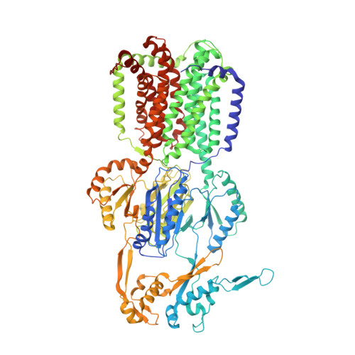
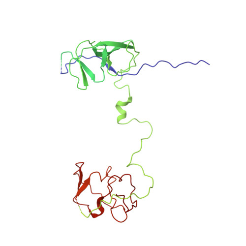
6VEJ

TriABC transporter from Pseudomonas aeruginosa


Experimental Data Snapshot

  • Method: ELECTRON MICROSCOPY
  • Resolution: 4.50 Å
  • Aggregation State: PARTICLE 
  • Reconstruction Method: SINGLE PARTICLE 

Starting Models: experimental
View more details

wwPDB Validation   3D Report Full Report


This is version 1.3 of the entry. See complete history


Literature

A "Drug Sweeping" State of the TriABC Triclosan Efflux Pump from Pseudomonas aeruginosa.

Fabre, L.Ntreh, A.T.Yazidi, A.Leus, I.V.Weeks, J.W.Bhattacharyya, S.Ruickoldt, J.Rouiller, I.Zgurskaya, H.I.Sygusch, J.

(2021) Structure 29: 261

  • DOI: https://doi.org/10.1016/j.str.2020.09.001
  • Primary Citation of Related Structures:  
    6VEJ

  • PubMed Abstract: 

    The structure of the TriABC inner membrane component of the triclosan/SDS-specific efflux pump from Pseudomonas aeruginosa was determined by cryoelectron microscopy to 4.5 Å resolution. The complete structure of the inner membrane transporter TriC of the resistance-nodulation-division (RND) superfamily was solved, including a partial structure of the fused periplasmic membrane fusion subunits, TriA and TriB. The substrate-free conformation of TriABC represents an intermediate step in efflux complex assembly before the engagement of the outer membrane channel. Structural analysis identified a tunnel network whose constriction impedes substrate efflux, indicating inhibition of TriABC in the unengaged state. Blind docking studies revealed binding to TriC at the same loci by substrates and bulkier non-substrates. Together with functional analyses, we propose that selective substrate translocation involves conformational gating at the tunnel narrowing that, together with conformational ordering of TriA and TriB, creates an engaged state capable of mediating substrate efflux.


  • Organizational Affiliation

    McGill University, Department of Anatomy and Cell Biology, Montreal, QC H3A 0G4, Canada.


Macromolecules
Find similar proteins by:  (by identity cutoff)  |  3D Structure
Entity ID: 1
MoleculeChains Sequence LengthOrganismDetailsImage
Probable Resistance-Nodulation-Cell Division (RND) efflux transporter
A, B, C
1,022Pseudomonas aeruginosaMutation(s): 0 
Gene Names: PA0158
Membrane Entity: Yes 
UniProt
Find proteins for Q9I6X4 (Pseudomonas aeruginosa (strain ATCC 15692 / DSM 22644 / CIP 104116 / JCM 14847 / LMG 12228 / 1C / PRS 101 / PAO1))
Explore Q9I6X4 
Go to UniProtKB:  Q9I6X4
Entity Groups  
Sequence Clusters30% Identity50% Identity70% Identity90% Identity95% Identity100% Identity
UniProt GroupQ9I6X4
Sequence Annotations
Expand
  • Reference Sequence
Find similar proteins by:  (by identity cutoff)  |  3D Structure
Entity ID: 2
MoleculeChains Sequence LengthOrganismDetailsImage
Probable Resistance-Nodulation-Cell Division (RND) efflux membrane fusion protein,Probable Resistance-Nodulation-Cell Division (RND) efflux membrane fusion proteinD [auth P],
E [auth Q],
F [auth R]
695Pseudomonas aeruginosaMutation(s): 0 
Gene Names: PA0156PA0157
UniProt
Find proteins for Q9I6X6 (Pseudomonas aeruginosa (strain ATCC 15692 / DSM 22644 / CIP 104116 / JCM 14847 / LMG 12228 / 1C / PRS 101 / PAO1))
Explore Q9I6X6 
Go to UniProtKB:  Q9I6X6
Find proteins for Q9I6X5 (Pseudomonas aeruginosa (strain ATCC 15692 / DSM 22644 / CIP 104116 / JCM 14847 / LMG 12228 / 1C / PRS 101 / PAO1))
Explore Q9I6X5 
Go to UniProtKB:  Q9I6X5
Entity Groups  
Sequence Clusters30% Identity50% Identity70% Identity90% Identity95% Identity100% Identity
UniProt GroupsQ9I6X5Q9I6X6
Sequence Annotations
Expand
  • Reference Sequence
Small Molecules
Ligands 3 Unique
IDChains Name / Formula / InChI Key2D Diagram3D Interactions
E2V
Query on E2V

Download Ideal Coordinates CCD File 
J [auth P],
L [auth Q],
N [auth R]
(2R)-2-(hexadecanoyloxy)propyl nonadecanoate
C38 H74 O4
AZEJVFRTMDKJQJ-PSXMRANNSA-N
LMU
Query on LMU

Download Ideal Coordinates CCD File 
G [auth A],
H [auth B],
I [auth C]
DODECYL-ALPHA-D-MALTOSIDE
C24 H46 O11
NLEBIOOXCVAHBD-YHBSTRCHSA-N
PLM
Query on PLM

Download Ideal Coordinates CCD File 
K [auth P],
M [auth Q],
O [auth R]
PALMITIC ACID
C16 H32 O2
IPCSVZSSVZVIGE-UHFFFAOYSA-N
Experimental Data & Validation

Experimental Data

  • Method: ELECTRON MICROSCOPY
  • Resolution: 4.50 Å
  • Aggregation State: PARTICLE 
  • Reconstruction Method: SINGLE PARTICLE 
EM Software:
TaskSoftware PackageVersion
RECONSTRUCTIONRELION1.3

Structure Validation

View Full Validation Report


Entry History & Funding Information

Deposition Data


Funding OrganizationLocationGrant Number
Other governmentCanadaFRSQ- GEPROM- Seed Grant
Natural Sciences and Engineering Research Council (NSERC, Canada)CanadaRGPIN-2014-04798
Natural Sciences and Engineering Research Council (NSERC, Canada)CanadaRGPIN-2016-04898
Canadian Institutes of Health Research (CIHR)CanadaMOP 86693
National Institutes of Health/National Institute Of Allergy and Infectious Diseases (NIH/NIAID)United StatesAI052293
Other privateAustraliaUniversity of Melbourne- Start-up Funds

Revision History  (Full details and data files)

  • Version 1.0: 2020-09-30
    Type: Initial release
  • Version 1.1: 2020-10-28
    Changes: Database references
  • Version 1.2: 2021-03-17
    Changes: Database references
  • Version 1.3: 2024-10-23
    Changes: Data collection, Database references, Refinement description, Structure summary