6VC8 | pdb_00006vc8

Crystal structure of wild-type KRAS4b(1-169) in complex with GMPPNP and Mg ion


Experimental Data Snapshot

  • Method: X-RAY DIFFRACTION
  • Resolution: 2.50 Å
  • R-Value Free: 
    0.277 (Depositor), 0.271 (DCC) 
  • R-Value Work: 
    0.212 (Depositor), 0.214 (DCC) 
  • R-Value Observed: 
    0.215 (Depositor) 

Starting Model: experimental
View more details

wwPDB Validation 3D Report Full Report

Validation slider image for 6VC8

Ligand Structure Quality Assessment 


This is version 1.2 of the entry. See complete history

Literature

Machine learning-driven multiscale modeling reveals lipid-dependent dynamics of RAS signaling proteins.

Ingolfsson, H.I.Neale, C.Carpenter, T.S.Shrestha, R.Lopez, C.A.Tran, T.H.Oppelstrup, T.Bhatia, H.Stanton, L.G.Zhang, X.Sundram, S.Di Natale, F.Agarwal, A.Dharuman, G.Kokkila Schumacher, S.I.L.Turbyville, T.Gulten, G.Van, Q.N.Goswami, D.Jean-Francois, F.Agamasu, C.Hettige, J.J.Travers, T.Sarkar, S.Surh, M.P.Yang, Y.Moody, A.Liu, S.Van Essen, B.C.Voter, A.F.Ramanathan, A.Hengartner, N.W.Simanshu, D.K.Stephen, A.G.Bremer, P.T.Gnanakaran, S.Glosli, J.N.Lightstone, F.C.McCormick, F.Nissley, D.V.Streitz, F.H.

(2022) Proc Natl Acad Sci U S A 119

  • DOI: https://doi.org/10.1073/pnas.2113297119
  • Primary Citation Related Structures: 
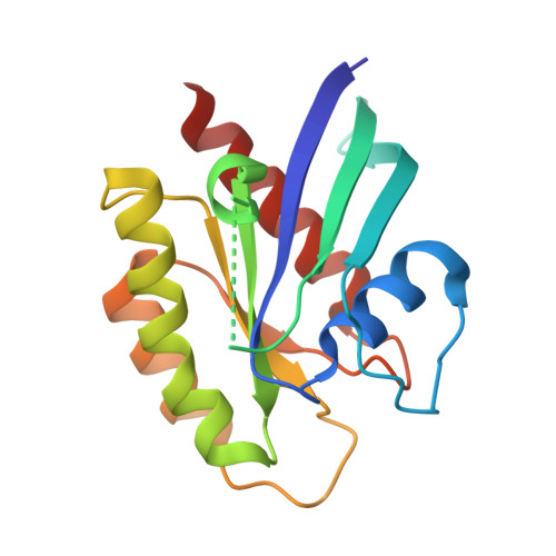
    6VC8

  • PubMed Abstract: 

    RAS is a signaling protein associated with the cell membrane that is mutated in up to 30% of human cancers. RAS signaling has been proposed to be regulated by dynamic heterogeneity of the cell membrane. Investigating such a mechanism requires near-atomistic detail at macroscopic temporal and spatial scales, which is not possible with conventional computational or experimental techniques. We demonstrate here a multiscale simulation infrastructure that uses machine learning to create a scale-bridging ensemble of over 100,000 simulations of active wild-type KRAS on a complex, asymmetric membrane. Initialized and validated with experimental data (including a new structure of active wild-type KRAS), these simulations represent a substantial advance in the ability to characterize RAS-membrane biology. We report distinctive patterns of local lipid composition that correlate with interfacially promiscuous RAS multimerization. These lipid fingerprints are coupled to RAS dynamics, predicted to influence effector binding, and therefore may be a mechanism for regulating cell signaling cascades.


  • Organizational Affiliation
    • Physical and Life Sciences Directorate, Lawrence Livermore National Laboratory, Livermore, CA 94550.

Macromolecule Content 

  • Total Structure Weight: 59.63 kDa 
  • Atom Count: 3,689 
  • Modeled Residue Count: 470 
  • Deposited Residue Count: 510 
  • Unique protein chains: 1

Macromolecules

Find similar proteins by:|  3D Structure
Entity ID: 1
MoleculeChains  Sequence LengthOrganismDetailsImage
GTPase KRas
A, B, C
170Homo sapiensMutation(s): 0 
Gene Names: KRASKRAS2RASK2
EC: 3.6.5.2
UniProt & NIH Common Fund Data Resources
Find proteins for P01116 (Homo sapiens)
Explore P01116 
Go to UniProtKB:  P01116
PHAROS:  P01116
GTEx:  ENSG00000133703 
Entity Groups
Sequence Clusters30% Identity50% Identity70% Identity90% Identity95% Identity100% Identity
UniProt GroupP01116
Sequence Annotations
Expand
Reference Sequence

Experimental Data & Validation

Experimental Data

  • Method: X-RAY DIFFRACTION
  • Resolution: 2.50 Å
  • R-Value Free:  0.277 (Depositor), 0.271 (DCC) 
  • R-Value Work:  0.212 (Depositor), 0.214 (DCC) 
  • R-Value Observed: 0.215 (Depositor) 
Space Group: C 1 2 1
Unit Cell:
Length ( Å )Angle ( ˚ )
a = 69.21α = 90
b = 82.33β = 112.92
c = 88.14γ = 90
Software Package:
Software NamePurpose
XDSdata reduction
XSCALEdata scaling
PHASERphasing
PHENIXrefinement
PDB_EXTRACTdata extraction

Structure Validation

View Full Validation Report


Ligand Structure Quality Assessment 


Entry History 

& Funding Information

Deposition Data


Funding OrganizationLocationGrant Number
National Institutes of Health/National Cancer Institute (NIH/NCI)United States--

Revision History  (Full details and data files)

  • Version 1.0: 2021-02-10
    Type: Initial release
  • Version 1.1: 2022-01-12
    Changes: Database references
  • Version 1.2: 2023-10-11
    Changes: Data collection, Refinement description