6VAV | pdb_00006vav

Peanut lectin complexed with divalent N-beta-D-galactopyranosyl-L-succinamoyl derivative (diNGS)


Experimental Data Snapshot

  • Method: X-RAY DIFFRACTION
  • Resolution: 1.85 Å
  • R-Value Free: 
    0.225 (Depositor), 0.228 (DCC) 
  • R-Value Work: 
    0.205 (Depositor), 0.208 (DCC) 
  • R-Value Observed: 
    0.206 (Depositor) 

Starting Model: experimental
View more details

wwPDB Validation   3D Report Full Report


Ligand Structure Quality Assessment 


This is version 1.2 of the entry. See complete history


Literature

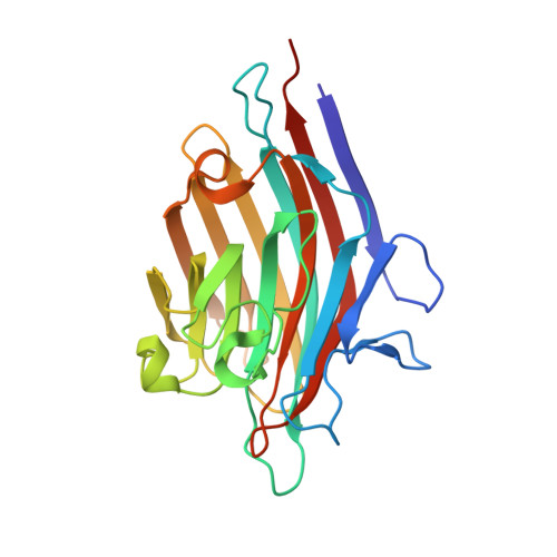
Crystal structures of peanut lectin in the presence of synthetic beta-N- and beta-S-galactosides disclose evidence for the recognition of different glycomimetic ligands.

Cagnoni, A.J.Primo, E.D.Klinke, S.Cano, M.E.Giordano, W.Marino, K.V.Kovensky, J.Goldbaum, F.A.Uhrig, M.L.Otero, L.H.

(2020) Acta Crystallogr D Struct Biol 76: 1080-1091

  • DOI: https://doi.org/10.1107/S2059798320012371
  • Primary Citation Related Structures: 
    6V95, 6VAV, 6VAW, 6VC3, 6VC4, 6VGF

  • PubMed Abstract: 

    Carbohydrate-lectin interactions are involved in important cellular recognition processes, including viral and bacterial infections, inflammation and tumor metastasis. Hence, structural studies of lectin-synthetic glycan complexes are essential for understanding lectin-recognition processes and for the further design of promising chemotherapeutics that interfere with sugar-lectin interactions. Plant lectins are excellent models for the study of the molecular-recognition process. Among them, peanut lectin (PNA) is highly relevant in the field of glycobiology because of its specificity for β-galactosides, showing high affinity towards the Thomsen-Friedenreich antigen, a well known tumor-associated carbohydrate antigen. Given this specificity, PNA is one of the most frequently used molecular probes for the recognition of tumor cell-surface O-glycans. Thus, it has been extensively used in glycobiology for inhibition studies with a variety of β-galactoside and β-lactoside ligands. Here, crystal structures of PNA are reported in complex with six novel synthetic hydrolytically stable β-N- and β-S-galactosides. These complexes disclosed key molecular-binding interactions of the different sugars with PNA at the atomic level, revealing the roles of specific water molecules in protein-ligand recognition. Furthermore, binding-affinity studies by isothermal titration calorimetry showed dissociation-constant values in the micromolar range, as well as a positive multivalency effect in terms of affinity in the case of the divalent compounds. Taken together, this work provides a qualitative structural rationale for the upcoming synthesis of optimized glycoclusters designed for the study of lectin-mediated biological processes. The understanding of the recognition of β-N- and β-S-galactosides by PNA represents a benchmark in protein-carbohydrate interactions since they are novel synthetic ligands that do not belong to the family of O-linked glycosides.


  • Organizational Affiliation
    • Laboratorio de Glicómica Funcional y Molecular, Instituto de Biología y Medicina Experimental, IBYME-CONICET, Vuelta de Obligado 2490, C1428ADN Buenos Aires, Argentina.

Macromolecules
Find similar proteins by:  (by identity cutoff)  |  3D Structure
Entity ID: 1
MoleculeChains Sequence LengthOrganismDetailsImage
Galactose-binding lectin
A, B, C, D
236Arachis hypogaeaMutation(s): 0 
UniProt
Find proteins for P02872 (Arachis hypogaea)
Explore P02872 
Go to UniProtKB:  P02872
Entity Groups  
Sequence Clusters30% Identity50% Identity70% Identity90% Identity95% Identity100% Identity
UniProt GroupP02872
Sequence Annotations
Expand
  • Reference Sequence
Small Molecules
Ligands 3 Unique
IDChains Name / Formula / InChI Key2D Diagram3D Interactions
QTY
Query on QTY

Download Ideal Coordinates CCD File 
E [auth A],
H [auth B],
K [auth C],
L [auth D]
N-[[1-[(3S,3aR,6S,6aR)-6-[4-[[[4-[[(2R,3R,4S,5R,6R)-6-(hydroxymethyl)-3,4,5-tris(oxidanyl)oxan-2-yl]amino]-4-oxidanylidene-butanoyl]amino]methyl]-1,2,3-triazol-1-yl]-2,3,3a,5,6,6a-hexahydrofuro[3,2-b]furan-3-yl]-1,2,3-triazol-4-yl]methyl]-N'-[(2R,3R,4S,5R,6R)-6-(hydroxymethyl)-3,4,5-tris(oxidanyl)oxan-2-yl]butanediamide
C32 H48 N10 O16
WUNJKQMXDITVGU-ADJUJXGJSA-N
MN
Query on MN

Download Ideal Coordinates CCD File 
F [auth A],
I [auth B],
M [auth D]
MANGANESE (II) ION
Mn
WAEMQWOKJMHJLA-UHFFFAOYSA-N
CA
Query on CA

Download Ideal Coordinates CCD File 
G [auth A],
J [auth B],
N [auth D]
CALCIUM ION
Ca
BHPQYMZQTOCNFJ-UHFFFAOYSA-N
Experimental Data & Validation

Experimental Data

  • Method: X-RAY DIFFRACTION
  • Resolution: 1.85 Å
  • R-Value Free:  0.225 (Depositor), 0.228 (DCC) 
  • R-Value Work:  0.205 (Depositor), 0.208 (DCC) 
  • R-Value Observed: 0.206 (Depositor) 
Space Group: P 2 21 21
Unit Cell:
Length ( Å )Angle ( ˚ )
a = 75.76α = 90
b = 124.64β = 90
c = 128.375γ = 90
Software Package:
Software NamePurpose
BUSTERrefinement
PDB_EXTRACTdata extraction
XDSdata reduction
Aimlessdata scaling
PHASERphasing
MxCuBEdata collection

Structure Validation

View Full Validation Report


Ligand Structure Quality Assessment 


Entry History & Funding Information

Deposition Data


Funding OrganizationLocationGrant Number
National Research Council (NRC, Argentina)ArgentinaPICT-2015-0621
National Research Council (NRC, Argentina)ArgentinaPICT-2016-1425

Revision History  (Full details and data files)

  • Version 1.0: 2020-10-28
    Type: Initial release
  • Version 1.1: 2020-11-18
    Changes: Database references
  • Version 1.2: 2023-10-11
    Changes: Advisory, Data collection, Database references, Refinement description