6VAB | pdb_00006vab

Mouse retromer sub-structure: VPS35/VPS35 flat dimer


Experimental Data Snapshot

  • Method: ELECTRON MICROSCOPY
  • Resolution: 4.90 Å
  • Aggregation State: PARTICLE 
  • Reconstruction Method: SINGLE PARTICLE 

wwPDB Validation 3D Report Full Report

Validation slider image for 6VAB

This is version 1.2 of the entry. See complete history

Literature

Mammalian Retromer Is an Adaptable Scaffold for Cargo Sorting from Endosomes.

Kendall, A.K.Xie, B.Xu, P.Wang, J.Burcham, R.Frazier, M.N.Binshtein, E.Wei, H.Graham, T.R.Nakagawa, T.Jackson, L.P.

(2020) Structure 28: 393-405.e4

  • DOI: https://doi.org/10.1016/j.str.2020.01.009
  • Primary Citation Related Structures: 
    6VAB, 6VAC

  • PubMed Abstract: 

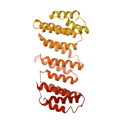
    Metazoan retromer (VPS26/VPS35/VPS29) associates with sorting nexins on endosomal tubules to sort proteins to the trans-Golgi network or plasma membrane. Mechanisms of metazoan retromer assembly remain undefined. We combine single-particle cryoelectron microscopy with biophysical methods to uncover multiple oligomer structures. 2D class averages reveal mammalian heterotrimers; dimers of trimers; tetramers of trimers; and flat chains. These species are further supported by biophysical solution studies. We provide reconstructions of all species, including key sub-structures (∼5 Å resolution). Local resolution variation suggests that heterotrimers and dimers adopt multiple conformations. Our structures identify a flexible, highly conserved electrostatic dimeric interface formed by VPS35 subunits. We generate structure-based mutants to disrupt this interface in vitro. Equivalent mutations in yeast demonstrate a mild cargo-sorting defect. Our data suggest the metazoan retromer is an adaptable and plastic scaffold that accommodates interactions with different sorting nexins to sort multiple cargoes from endosomes their final destinations.


  • Organizational Affiliation
    • Department of Biological Sciences, Vanderbilt University, Nashville, TN 37232, USA; Center for Structural Biology, Vanderbilt University, Nashville, TN 37232, USA.

Macromolecule Content 

  • Total Structure Weight: 224.69 kDa 
  • Atom Count: 4,642 
  • Modeled Residue Count: 938 
  • Deposited Residue Count: 1,956 
  • Unique protein chains: 2

Macromolecules

Find similar proteins by:|  3D Structure
Entity ID: 1
MoleculeChains  Sequence LengthOrganismDetailsImage
Vacuolar protein sorting-associated protein 29
A, C
182Mus musculusMutation(s): 0 
Gene Names: Vps29
UniProt & NIH Common Fund Data Resources
Find proteins for Q9QZ88 (Mus musculus)
Explore Q9QZ88 
Go to UniProtKB:  Q9QZ88
IMPC:  MGI:1928344
Entity Groups
Sequence Clusters30% Identity50% Identity70% Identity90% Identity95% Identity100% Identity
UniProt GroupQ9QZ88
Sequence Annotations
Expand
Reference Sequence
Find similar proteins by:|  3D Structure
Entity ID: 2
MoleculeChains  Sequence LengthOrganismDetailsImage
Vacuolar protein sorting-associated protein 35
B, D
796Mus musculusMutation(s): 0 
Gene Names: Vps35Mem3
UniProt & NIH Common Fund Data Resources
Find proteins for Q9EQH3 (Mus musculus)
Explore Q9EQH3 
Go to UniProtKB:  Q9EQH3
IMPC:  MGI:1890467
Entity Groups
Sequence Clusters30% Identity50% Identity70% Identity90% Identity95% Identity100% Identity
UniProt GroupQ9EQH3
Sequence Annotations
Expand
Reference Sequence

Experimental Data & Validation

Experimental Data

  • Method: ELECTRON MICROSCOPY
  • Resolution: 4.90 Å
  • Aggregation State: PARTICLE 
  • Reconstruction Method: SINGLE PARTICLE 

Structure Validation

View Full Validation Report


Entry History 

& Funding Information

Deposition Data


Funding OrganizationLocationGrant Number
National Institutes of Health/National Institute of General Medical Sciences (NIH/NIGMS)--

Revision History  (Full details and data files)

  • Version 1.0: 2020-02-19
    Type: Initial release
  • Version 1.1: 2020-04-15
    Changes: Database references
  • Version 1.2: 2024-03-06
    Changes: Data collection, Database references