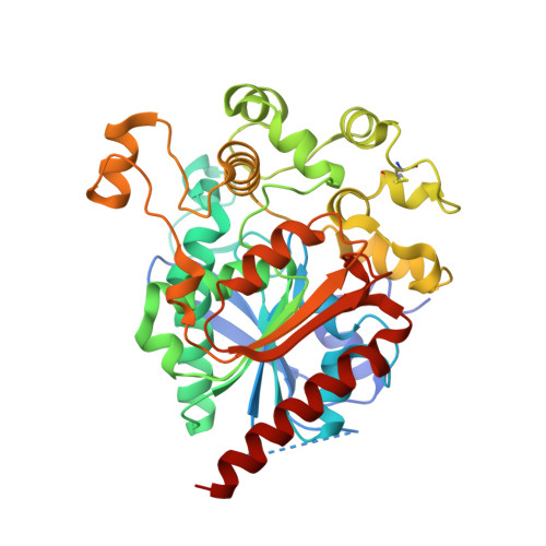
6V7N

Crystal Structure of a human Lysosome Resident Glycoprotein, Lysosomal Acid Lipase, and its Implications in Cholesteryl Ester Storage Disease (CESD)


Experimental Data Snapshot

  • Method: X-RAY DIFFRACTION
  • Resolution: 2.62 Å
  • R-Value Free: 0.222 
  • R-Value Work: 0.185 
  • R-Value Observed: 0.187 

Starting Model: experimental
View more details

wwPDB Validation   3D Report Full Report

Currently 6V7N does not have a validation slider image.


This is version 2.3 of the entry. See complete history


Literature

Crystal structure of human lysosomal acid lipase and its implications in cholesteryl ester storage disease.

Rajamohan, F.Reyes, A.R.Tu, M.Nedoma, N.L.Hoth, L.R.Schwaid, A.G.Kurumbail, R.G.Ward, J.Han, S.

(2020) J Lipid Res 61: 1192-1202

  • DOI: https://doi.org/10.1194/jlr.RA120000748
  • Primary Citation of Related Structures:  
    6V7N

  • PubMed Abstract: 

    Lysosomal acid lipase (LAL) is a serine hydrolase that hydrolyzes cholesteryl ester (CE) and TGs delivered to the lysosomes into free cholesterol and fatty acids. LAL deficiency due to mutations in the LAL gene ( LIPA ) results in accumulation of TGs and cholesterol esters in various tissues of the body leading to pathological conditions such as Wolman's disease and CE storage disease (CESD). Here, we present the first crystal structure of recombinant human LAL (HLAL) to 2.6 Å resolution in its closed form. The crystal structure was enabled by mutating three of the six potential glycosylation sites. The overall structure of HLAL closely resembles that of the evolutionarily related human gastric lipase (HGL). It consists of a core domain belonging to the classical α/β hydrolase-fold family with a classical catalytic triad (Ser-153, His-353, Asp-324), an oxyanion hole, and a "cap" domain, which regulates substrate entry to the catalytic site. Most significant structural differences between HLAL and HGL exist at the lid region. Deletion of the short helix, 238 NLCFLLC 244 , at the lid region implied a possible role in regulating the highly hydrophobic substrate binding site from self-oligomerization during interfacial activation. We also performed molecular dynamic simulations of dog gastric lipase (lid-open form) and HLAL to gain insights and speculated a possible role of the human mutant, H274Y, leading to CESD.


  • Organizational Affiliation

    Pfizer Worldwide Research, Groton, CT 06340 francis.rajamohan@pfizer.com.


Macromolecules
Find similar proteins by:  (by identity cutoff)  |  3D Structure
Entity ID: 1
MoleculeChains Sequence LengthOrganismDetailsImage
Lysosomal acid lipase/cholesteryl ester hydrolase
A, B
384Homo sapiensMutation(s): 3 
Gene Names: LIPA
EC: 3.1.1.13
Membrane Entity: Yes 
UniProt & NIH Common Fund Data Resources
Find proteins for P38571 (Homo sapiens)
Explore P38571 
Go to UniProtKB:  P38571
PHAROS:  P38571
GTEx:  ENSG00000107798 
Entity Groups  
Sequence Clusters30% Identity50% Identity70% Identity90% Identity95% Identity100% Identity
UniProt GroupP38571
Glycosylation
Glycosylation Sites: 2Go to GlyGen: P38571-1
Sequence Annotations
Expand
  • Reference Sequence
Oligosaccharides

Help

Entity ID: 2
MoleculeChains Length2D Diagram Glycosylation3D Interactions
2-acetamido-2-deoxy-beta-D-glucopyranose-(1-4)-2-acetamido-2-deoxy-beta-D-glucopyranose
C, D
2N-Glycosylation
Glycosylation Resources
GlyTouCan:  G42666HT
GlyCosmos:  G42666HT
GlyGen:  G42666HT
Experimental Data & Validation

Experimental Data

  • Method: X-RAY DIFFRACTION
  • Resolution: 2.62 Å
  • R-Value Free: 0.222 
  • R-Value Work: 0.185 
  • R-Value Observed: 0.187 
  • Space Group: P 21 21 2
Unit Cell:
Length ( Å )Angle ( ˚ )
a = 97.145α = 90
b = 165.486β = 90
c = 60.36γ = 90
Software Package:
Software NamePurpose
BUSTERrefinement
PDB_EXTRACTdata extraction
HKL-3000data reduction
SCALAdata scaling
BUSTERphasing

Structure Validation

View Full Validation Report

Currently 6V7N does not have a validation slider image.


Entry History 

Deposition Data

  • Released Date: 2020-06-17 
  • Deposition Author(s): Han, S.

Revision History  (Full details and data files)

  • Version 1.0: 2020-06-17
    Type: Initial release
  • Version 2.0: 2020-07-29
    Type: Remediation
    Reason: Carbohydrate remediation
    Changes: Atomic model, Data collection, Derived calculations, Structure summary
  • Version 2.1: 2020-08-12
    Changes: Database references, Structure summary
  • Version 2.2: 2023-10-11
    Changes: Data collection, Database references, Refinement description
  • Version 2.3: 2024-11-20
    Changes: Structure summary