


Crystal structure of human DNA polymerase eta complexed with N7-nitrogen half-mustard guanine (NHMG) and dCTP*
Jung, H., Lee, S.To be published.
Experimental Data Snapshot
Starting Model: experimental
View more details
Entity ID: 1 | |||||
|---|---|---|---|---|---|
| Molecule | Chains | Sequence Length | Organism | Details | Image |
| DNA polymerase eta | 432 | Homo sapiens | Mutation(s): 0 Gene Names: POLH, RAD30, RAD30A, XPV EC: 2.7.7.7 |  | |
UniProt & NIH Common Fund Data Resources | |||||
PHAROS: Q9Y253 GTEx: ENSG00000170734 | |||||
Entity Groups | |||||
| Sequence Clusters | 30% Identity50% Identity70% Identity90% Identity95% Identity100% Identity | ||||
| UniProt Group | Q9Y253 | ||||
Sequence AnnotationsExpand | |||||
Reference Sequence | |||||
Entity ID: 2 | ||||
| Molecule | Chains | Length | Organism | Image |
|---|---|---|---|---|
| DNA (5'-D(*AP*GP*TP*GP*TP*GP*AP*G)-3') | B [auth P] | 8 | Homo sapiens | 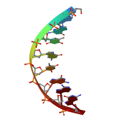 |
Sequence AnnotationsExpand | ||||
Reference Sequence | ||||
Entity ID: 3 | ||||
| Molecule | Chains | Length | Organism | Image |
|---|---|---|---|---|
| DNA (5'-D(*CP*AP*T*(QRV)P*CP*TP*CP*AP*CP*AP*CP*T)-3') | C [auth T] | 12 | Homo sapiens | 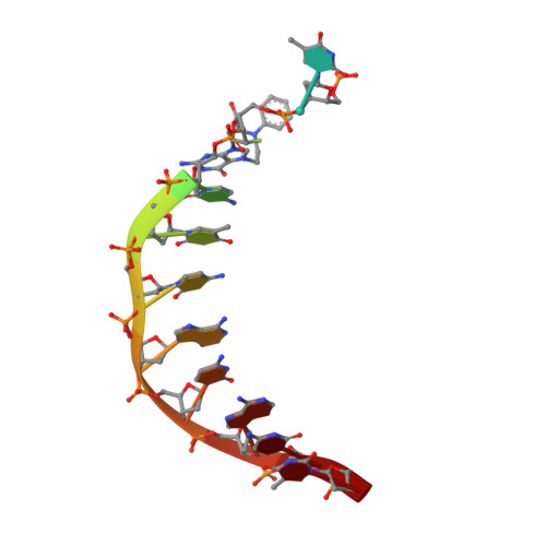 |
Sequence AnnotationsExpand | ||||
Reference Sequence | ||||
| Ligands 2 Unique | |||||
|---|---|---|---|---|---|
| ID | Chains | Name / Formula / InChI Key | 2D Diagram | 3D Interactions | |
| 0KX Download:Ideal Coordinates CCD File | D [auth A] | 2'-deoxy-5'-O-[(R)-hydroxy{[(R)-hydroxy(phosphonooxy)phosphoryl]amino}phosphoryl]cytidine C9 H17 N4 O12 P3 STYMTWKSQLVXJN-SHYZEUOFSA-N |  | ||
| MN Download:Ideal Coordinates CCD File | E [auth A] | MANGANESE (II) ION Mn WAEMQWOKJMHJLA-UHFFFAOYSA-N |  | ||
| Length ( Å ) | Angle ( ˚ ) |
|---|---|
| a = 98.911 | α = 90 |
| b = 98.911 | β = 90 |
| c = 82.138 | γ = 120 |
| Software Name | Purpose |
|---|---|
| PHENIX | refinement |
| HKL-2000 | data reduction |
| HKL-2000 | data scaling |
| MOLREP | phasing |


