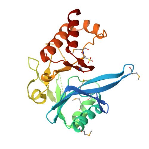
Crystal Structure of the NDM_FIM-1 like Metallo-beta-Lactamase from Erythrobacter litoralis in the Mono-Zinc Form
Kim, Y., Maltseva, N., Mulligan, R., Endres, M., Joachimiak, A., Center for Structural Genomics of Infectious Diseases (CSGID)To be published.