6V0N

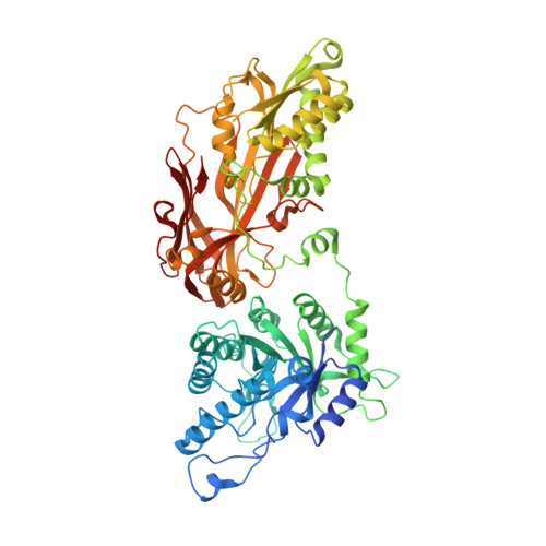
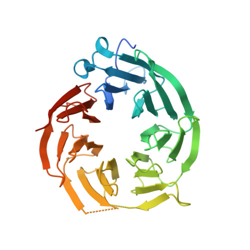
PRMT5 bound to PBM peptide from Riok1


Experimental Data Snapshot

  • Method: X-RAY DIFFRACTION
  • Resolution: 2.11 Å
  • R-Value Free: 0.235 
  • R-Value Work: 0.203 
  • R-Value Observed: 0.204 

Starting Model: experimental
View more details

wwPDB Validation   3D Report Full Report

Currently 6V0N does not have a validation slider image.


This is version 1.4 of the entry. See complete history


Literature

Molecular basis for substrate recruitment to the PRMT5 methylosome.

Mulvaney, K.M.Blomquist, C.Acharya, N.Li, R.Ranaghan, M.J.O'Keefe, M.Rodriguez, D.J.Young, M.J.Kesar, D.Pal, D.Stokes, M.Nelson, A.J.Jain, S.S.Yang, A.Mullin-Bernstein, Z.Columbus, J.Bozal, F.K.Skepner, A.Raymond, D.LaRussa, S.McKinney, D.C.Freyzon, Y.Baidi, Y.Porter, D.Aguirre, A.J.Ianari, A.McMillan, B.Sellers, W.R.

(2021) Mol Cell 81: 3481

  • DOI: https://doi.org/10.1016/j.molcel.2021.07.019
  • Primary Citation of Related Structures:  
    6V0N, 6V0O

  • PubMed Abstract: 

    PRMT5 is an essential arginine methyltransferase and a therapeutic target in MTAP-null cancers. PRMT5 uses adaptor proteins for substrate recruitment through a previously undefined mechanism. Here, we identify an evolutionarily conserved peptide sequence shared among the three known substrate adaptors (CLNS1A, RIOK1, and COPR5) and show that it is necessary and sufficient for interaction with PRMT5. We demonstrate that PRMT5 uses modular adaptor proteins containing a common binding motif for substrate recruitment, comparable with other enzyme classes such as kinases and E3 ligases. We structurally resolve the interface with PRMT5 and show via genetic perturbation that it is required for methylation of adaptor-recruited substrates including the spliceosome, histones, and ribosomal complexes. Furthermore, disruption of this site affects Sm spliceosome activity, leading to intron retention. Genetic disruption of the PRMT5-substrate adaptor interface impairs growth of MTAP-null tumor cells and is thus a site for development of therapeutic inhibitors of PRMT5.


  • Organizational Affiliation

    Broad Institute, Cambridge, MA, USA.


Macromolecules
Find similar proteins by:  (by identity cutoff)  |  3D Structure
Entity ID: 1
MoleculeChains Sequence LengthOrganismDetailsImage
Protein arginine N-methyltransferase 5637Homo sapiensMutation(s): 0 
Gene Names: PRMT5HRMT1L5IBP72JBP1SKB1
EC: 2.1.1.320
UniProt & NIH Common Fund Data Resources
Find proteins for O14744 (Homo sapiens)
Explore O14744 
Go to UniProtKB:  O14744
PHAROS:  O14744
GTEx:  ENSG00000100462 
Entity Groups  
Sequence Clusters30% Identity50% Identity70% Identity90% Identity95% Identity100% Identity
UniProt GroupO14744
Sequence Annotations
Expand
  • Reference Sequence
Find similar proteins by:  (by identity cutoff)  |  3D Structure
Entity ID: 2
MoleculeChains Sequence LengthOrganismDetailsImage
Methylosome protein 50342Homo sapiensMutation(s): 0 
Gene Names: WDR77MEP50WD45HKMT1069Nbla10071
UniProt & NIH Common Fund Data Resources
Find proteins for Q9BQA1 (Homo sapiens)
Explore Q9BQA1 
Go to UniProtKB:  Q9BQA1
PHAROS:  Q9BQA1
GTEx:  ENSG00000116455 
Entity Groups  
Sequence Clusters30% Identity50% Identity70% Identity90% Identity95% Identity100% Identity
UniProt GroupQ9BQA1
Sequence Annotations
Expand
  • Reference Sequence

Find similar proteins by:  Sequence   |   3D Structure  

Entity ID: 3
MoleculeChains Sequence LengthOrganismDetailsImage
Riok1 PBM peptide13Homo sapiensMutation(s): 0 
EC: 3.6.3 (UniProt), 2.7.11.1 (UniProt)
UniProt & NIH Common Fund Data Resources
Find proteins for Q9BRS2 (Homo sapiens)
Explore Q9BRS2 
Go to UniProtKB:  Q9BRS2
PHAROS:  Q9BRS2
GTEx:  ENSG00000124784 
Entity Groups  
Sequence Clusters30% Identity50% Identity70% Identity90% Identity95% Identity100% Identity
UniProt GroupQ9BRS2
Sequence Annotations
Expand
  • Reference Sequence
Small Molecules
Ligands 1 Unique
IDChains Name / Formula / InChI Key2D Diagram3D Interactions
SFG
Query on SFG

Download Ideal Coordinates CCD File 
D [auth A]SINEFUNGIN
C15 H23 N7 O5
LMXOHSDXUQEUSF-YECHIGJVSA-N
Binding Affinity Annotations 
IDSourceBinding Affinity
SFG BindingDB:  6V0N IC50: 8600 (nM) from 1 assay(s)
Experimental Data & Validation

Experimental Data

  • Method: X-RAY DIFFRACTION
  • Resolution: 2.11 Å
  • R-Value Free: 0.235 
  • R-Value Work: 0.203 
  • R-Value Observed: 0.204 
  • Space Group: I 2 2 2
Unit Cell:
Length ( Å )Angle ( ˚ )
a = 105.96α = 90
b = 139.41β = 90
c = 179.95γ = 90
Software Package:
Software NamePurpose
BUSTERrefinement
XDSdata reduction
XSCALEdata scaling
PHASERphasing
PDB_EXTRACTdata extraction

Structure Validation

View Full Validation Report

Currently 6V0N does not have a validation slider image.


Entry History 

Deposition Data

Revision History  (Full details and data files)

  • Version 1.0: 2020-08-26
    Type: Initial release
  • Version 1.1: 2020-09-02
    Changes: Database references
  • Version 1.2: 2021-08-25
    Changes: Database references
  • Version 1.3: 2021-09-15
    Changes: Database references
  • Version 1.4: 2023-10-11
    Changes: Data collection, Refinement description