6UZR | pdb_00006uzr

Crystal structure of GLUN1/GLUN2A ligand-binding domain in complex with glycine and homoquinolinic acid

  • Classification: MEMBRANE PROTEIN
  • Organism(s): Rattus norvegicus
  • Expression System: Escherichia coli BL21(DE3)
  • Mutation(s): No 

  • Deposited: 2019-11-15 Released: 2020-01-29 
  • Deposition Author(s): Wang, J.X., Furukawa, H.
  • Funding Organization(s): National Institutes of Health/National Institute of Neurological Disorders and Stroke (NIH/NINDS), National Institutes of Health/National Institute of Mental Health (NIH/NIMH)

Experimental Data Snapshot

  • Method: X-RAY DIFFRACTION
  • Resolution: 1.87 Å
  • R-Value Free: 
    0.207 (Depositor), 0.209 (DCC) 
  • R-Value Work: 
    0.173 (Depositor), 0.175 (DCC) 
  • R-Value Observed: 
    0.175 (Depositor) 

Starting Model: experimental
View more details

wwPDB Validation 3D Report Full Report

Validation slider image for 6UZR

Ligand Structure Quality Assessment 


This is version 1.3 of the entry. See complete history

Literature

Structural basis of subtype-selective competitive antagonism for GluN2C/2D-containing NMDA receptors.

Wang, J.X.Irvine, M.W.Burnell, E.S.Sapkota, K.Thatcher, R.J.Li, M.Simorowski, N.Volianskis, A.Collingridge, G.L.Monaghan, D.T.Jane, D.E.Furukawa, H.

(2020) Nat Commun 11: 423-423

  • DOI: https://doi.org/10.1038/s41467-020-14321-0
  • Primary Citation Related Structures: 
    6UZ6, 6UZG, 6UZR, 6UZW, 6UZX

  • PubMed Abstract: 

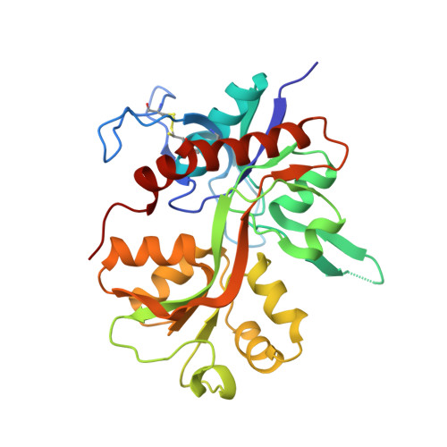
    N-Methyl-D-aspartate receptors (NMDARs) play critical roles in the central nervous system. Their heterotetrameric composition generates subtypes with distinct functional properties and spatio-temporal distribution in the brain, raising the possibility for subtype-specific targeting by pharmacological means for treatment of neurological diseases. While specific compounds for GluN2A and GluN2B-containing NMDARs are well established, those that target GluN2C and GluN2D are currently underdeveloped with low potency and uncharacterized binding modes. Here, using electrophysiology and X-ray crystallography, we show that UBP791 ((2S*,3R*)-1-(7-(2-carboxyethyl)phenanthrene-2-carbonyl)piperazine-2,3-dicarboxylic acid) inhibits GluN2C/2D with 40-fold selectivity over GluN2A-containing receptors, and that a methionine and a lysine residue in the ligand binding pocket (GluN2D-Met763/Lys766, GluN2C-Met736/Lys739) are the critical molecular elements for the subtype-specific binding. These findings led to development of UBP1700 ((2S*,3R*)-1-(7-(2-carboxyvinyl)phenanthrene-2-carbonyl)piperazine-2,3-dicarboxylic acid) which shows over 50-fold GluN2C/2D-selectivity over GluN2A with potencies in the low nanomolar range. Our study shows that the L-glutamate binding site can be targeted for GluN2C/2D-specific inhibition.


  • Organizational Affiliation
    • WM Keck Structural Biology Laboratory, Cold Spring Harbor Laboratory, Cold Spring Harbor, NY, 11724, USA.

Macromolecule Content 

  • Total Structure Weight: 65.57 kDa 
  • Atom Count: 4,987 
  • Modeled Residue Count: 566 
  • Deposited Residue Count: 574 
  • Unique protein chains: 2

Macromolecules

Find similar proteins by:|  3D Structure
Entity ID: 1
MoleculeChains  Sequence LengthOrganismDetailsImage
Glutamate receptor ionotropic, NMDA 1292Rattus norvegicusMutation(s): 0 
Gene Names: Grin1Nmdar1
UniProt
Find proteins for P35439 (Rattus norvegicus)
Explore P35439 
Go to UniProtKB:  P35439
Entity Groups
Sequence Clusters30% Identity50% Identity70% Identity90% Identity95% Identity100% Identity
UniProt GroupP35439
Sequence Annotations
Expand
Reference Sequence
Find similar proteins by:|  3D Structure
Entity ID: 2
MoleculeChains  Sequence LengthOrganismDetailsImage
Glutamate receptor ionotropic, NMDA 2A282Rattus norvegicusMutation(s): 0 
Gene Names: Grin2a
UniProt
Find proteins for Q00959 (Rattus norvegicus)
Explore Q00959 
Go to UniProtKB:  Q00959
Entity Groups
Sequence Clusters30% Identity50% Identity70% Identity90% Identity95% Identity100% Identity
UniProt GroupQ00959
Sequence Annotations
Expand
Reference Sequence

Experimental Data & Validation

Experimental Data

  • Method: X-RAY DIFFRACTION
  • Resolution: 1.87 Å
  • R-Value Free:  0.207 (Depositor), 0.209 (DCC) 
  • R-Value Work:  0.173 (Depositor), 0.175 (DCC) 
  • R-Value Observed: 0.175 (Depositor) 
Space Group: P 21 21 21
Unit Cell:
Length ( Å )Angle ( ˚ )
a = 54.367α = 90
b = 88.717β = 90
c = 125.483γ = 90
Software Package:
Software NamePurpose
autoPROCdata collection
XDSdata reduction
Aimlessdata scaling
pointlessdata scaling
REFMACphasing
PHENIXrefinement
Cootmodel building

Structure Validation

View Full Validation Report


Ligand Structure Quality Assessment 


Entry History 

& Funding Information

Deposition Data


Funding OrganizationLocationGrant Number
National Institutes of Health/National Institute of Neurological Disorders and Stroke (NIH/NINDS)United StatesNS111745
National Institutes of Health/National Institute of Mental Health (NIH/NIMH)United StatesMH085926

Revision History  (Full details and data files)

  • Version 1.0: 2020-01-29
    Type: Initial release
  • Version 1.1: 2020-02-05
    Changes: Database references
  • Version 1.2: 2023-10-11
    Changes: Advisory, Data collection, Database references, Refinement description
  • Version 1.3: 2024-10-23
    Changes: Structure summary